44 microscope labeled functions
Microscope Anatomy and function.ppt - Parts of a Compound... View Microscope Anatomy and function.ppt from BIO 120.32 at Post University. Parts of a Compound Microscope compound microscope Compound Microscope • A microscope is a very powerful. ... Ch 3 Microscope Anatomy 16-17.ppt. 30. microscopes lab review 117.ppt. Sinclair Community College. BIO MISC. Telescope; Modern Uses of Electron Microscopy for Detection of Viruses Specimens are flash-frozen in liquid nitrogen, transferred to the microscope in a special cold chamber, and viewed frozen in a special transmission electron microscope equipped with a cryo stage. Many digital images are made at different tilt angles and reconstructed by a computer into a three-dimensional representation (52, 73, 85, 86, 93 ...
Parts of Stereo Microscope (Dissecting microscope) - labeled diagram ... This type of microscope can provide a long working distance to accommodate larger objects and allow users to manipulate the objects under microscopic views. You may find many applications such as dissecting, micro-surgery, miniature manufacturing, and micro-engraving. This is why it is also named "Dissecting microscope".

Microscope labeled functions
Dissecting Microscope Parts And Functions. All You Need To Know Functions Of The Dissecting Microscope's Stand/Arm Head Support As previously mentioned, the stand supports the head of the microscope. If you consider the stand to be the microscope's spine, it allows the head to travel up and down to focus on the object in view. Focusing The coarse focus knob's location is on the rigid construction stand. Microscope Types (with labeled diagrams) and Functions Simple microscope labeled diagram Simple microscope functions It is used in industrial applications like: Watchmakers to assemble watches Cloth industry to count the number of threads or fibers in a cloth Jewelers to examine the finer parts of jewelry Miniature artists to examine and build their work Also used to inspect finer details on products PDF Parts of the Light Microscope - Science Spot B. NOSEPIECE microscope when carried Holds the HIGH- and LOW- power objective LENSES; can be rotated to change MAGNIFICATION. Power = 10 x 4 = 40 Power = 10 x 10 = 100 Power = 10 x 40 = 400 What happens as the power of magnification increases?
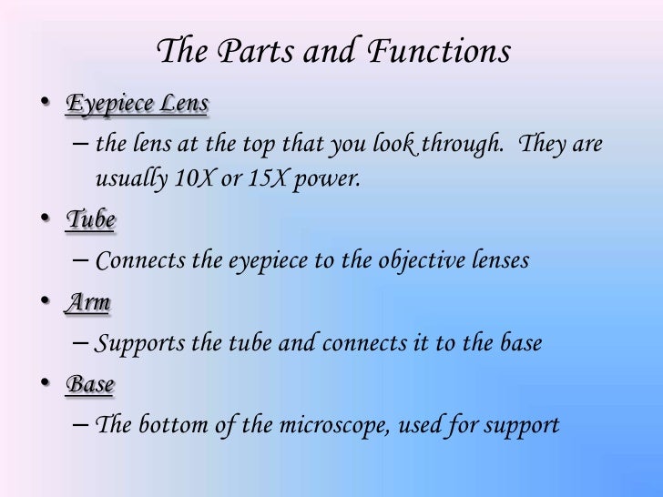
Microscope labeled functions. Parts of a Compound Microscope and Their Functions It controls the amount and intensity of light that enters the microscope. It can be either an iris diaphragm or a disc diaphragm. Condenser: It's a lense that's hidden beneath the stage. The size of the light beam is controlled by it. It collects and directs light from the mirror to the objective lens. Compound Microscope- Definition, Labeled Diagram, Principle, Parts, Uses The optical microscope often referred to as the light microscope, is a type of microscope that uses visible light and a system of lenses to magnify images of small subjects. There are two basic types of optical microscopes: Simple microscopes. Compound microscopes. The term "compound" in compound microscopes refers to the microscope having ... Microscope Parts & Specifications Microscope Parts & Specifications · The Functions & Parts of a Microscope · Eyepiece Lens: the lens at the top that you look through, usually 10x or 15x power. Parathyroid Gland Histology with Microscope Slide Image and Labeled ... The sample tissue section and diagram also show the numerous fat cells (adipose tissue). You may join anatomy learner on social media for a more updated labeled diagram on the parathyroid gland. Parathyroid gland microscope slide image drawing. This is a straightforward task to draw the microscope slide image of the parathyroid gland.
22 Parts Of a Microscope With Their Function And Labeled ... Parts Of a microscope. The main parts of a microscope that are easy to identify include: Head : The upper part of the microscope that houses the optical elements of the unit. Base: The base is attached to a frame (arm) that is connected to the head of the device. The base of the microscope provides stability to the device and allows the user ... Parts of the Microscope with Labeling (also Free Printouts) A standard microscope has two lenses namely the objective lens and the eyepiece. A light is needed to shine on the object and then reflected by the mirror into the lenses, hence, causing greater magnification. (1, 2, 3, and 4) Let us take a look at the different parts of microscopes and their respective functions. 1. Eyepiece Microscope Parts and Functions Flashcards | Quizlet regulates/controls the amount of light coming through the stage opening. Body Tube Maintains the proper distance between the eyepiece and the objective lens Stage Where you place the specimen that you want to view Stage Clips Holds the slides/specimen in place for viewing High Power objective Magnifies image 40X found on the nosepiece Base Microscope, Microscope Parts, Labeled Diagram, and Functions Microscopes are instruments that are used in science laboratories, to visualize very minute objects such as cells, microorganisms, giving a contrasting image, that is magnified. What is the Function of Microscope? A microscope is usually used for the study of microscopic algae, fungi, and biological specimens. What is Magnification?
Microscope Parts & Functions - AmScope Invented by a Dutch spectacle maker in the late 16th century, compound light microscopes use two sets of lenses to magnify images for study and observation. The first set of lenses are the oculars, or eyepieces, that the viewer looks into; the second set of lenses are the objectives, which are closest to the specimen. What are the parts of a microscope labeled? - SidmartinBio In this interactive, you can label the different parts of a microscope. What is the function of the arms of a microscope? It also carriers the microscopic illuminators. Arms - This is the part connecting the base and to the head and the eyepiece tube to the base of the microscope. Parts and Function of a Microscope Worksheets - DSoftSchools admin January 4, 2021. Some of the worksheets below are Parts and Function of a Microscope Worksheets with colorful charts and diagrams to help students familiarize with the parts of the microscope along with several important questions and activities with answers. Basic Instructions. Once you find your worksheet (s), you can either click on ... Compound Microscope Parts - Labeled Diagram and their Functions - Rs ... The information about an objective lens is labeled on its side. Key information that you should pay attention to is the magnification (i.e., 100x), NA (i.e., 1.25), and required media (i.e., Oil; no label means air). High-end microscopes also have achromatic, parcentered, or parfocal lenses.
microscope labeled parts and functions | Parts of a microscope with fu Optical parts and the functions The optical parts of the microscope are used to view, enlarge, and produce an image from a sample placed on a slide. These parts include Eyepiece: Eyepiece also contains ocular lens. It enhance the image of the viewer. This part is used for checking through the microscope. Eyepiece is found at the upper part of it.
Testis Histology - Complete Guide to Learn Histological ... Mar 01, 2021 · The functions of leydig cell of testis are – #1. Promotion of normal sexual behavior of animal #2. Enhance the growth and maintenance of the functions of pen-(is), male accessory sex organs and secondary sex characteristics #3. Control of spermatogenesis in animal testis. Testis histology labeled diagram
Animal Cell Diagram Under Light Microscope Labeled : Functions and Diagram Tuesday, April 20th 2021. | Diagram. Animal Cell Diagram Under Light Microscope. To make observations and draw scale. This shows a generalized animal cell under a light microscope. We all keep in mind that the human physique is amazingly elaborate and one way I discovered to comprehend it is by way of the style of human anatomy diagrams.
An Overview of the Cell Cycle - Molecular Biology of the Cell ... From the proportion of cells in such a population that are labeled (the labeling index), one can estimate the duration of S phase as a fraction of the whole cell cycle duration. Similarly, from the proportion of these cells in mitosis (the mitotic index ), one can estimate the duration of M phase .
PDF Microscope Parts and Functions - WPMU DEV Functions Microscope One or more lenses that makes an enlarged image of an object. 8/7/2018 2 •Simple •Compound •Stereoscopic •Electron Simple Microscope •Similar to a magnifying glass and has only one lens. 8/7/2018 3 Compound Microscope •Lets light pass through an object and
Parts of Microscope, Function, Names & Labeled Diagram Microscope parts labeled diagram gives us all the information about its parts and their position in the microscope. Microscope Parts Labeled Diagram The principle of the Microscope gives you an exact reason to use it. It works on the 3 principles. Magnification Resolving Power Numerical Aperture. Parts of Microscope Head Base Arm Eyepiece Lens
Microscope labeling and functions Flashcards | Quizlet Holds the low-power and high-power objective lenses; allows the lenses to rotate for viewing Revolving Nosepiece Magnifies about 4x Low-Power Objective Lens Magnify about 40x High-Power Objective Lenses Holds the slide in place Stage Clips Controls the amount of light passing through the opening of the stage Diaphragm
Compound Microscope Parts, Functions, and Labeled ... Parts of a Compound Microscope Each part of the compound microscope serves its own unique function, with each being important to the function of the scope as a whole. The individual parts of a compound microscope can vary heavily depending on the configuration & applications that the scope is being used for. Common compound microscope parts include: Compound Microscope Definitions for ...
Diagram Of Animal Cell Under Electron Microscope Labeled Diagram Of Animal Cell Under Electron Microscope. So, lets begin by drawing a rough-oval shape. It's a thin slice: Here's a diagram of a plant cell: The diagram is very clear, and labeled; but at the same time it is interpretive. We all do not forget that the human physique is quite problematic and a method I discovered to comprehend it is by ...


/images/library/67/Histology_Ileum.jpg)


Post a Comment for "44 microscope labeled functions"