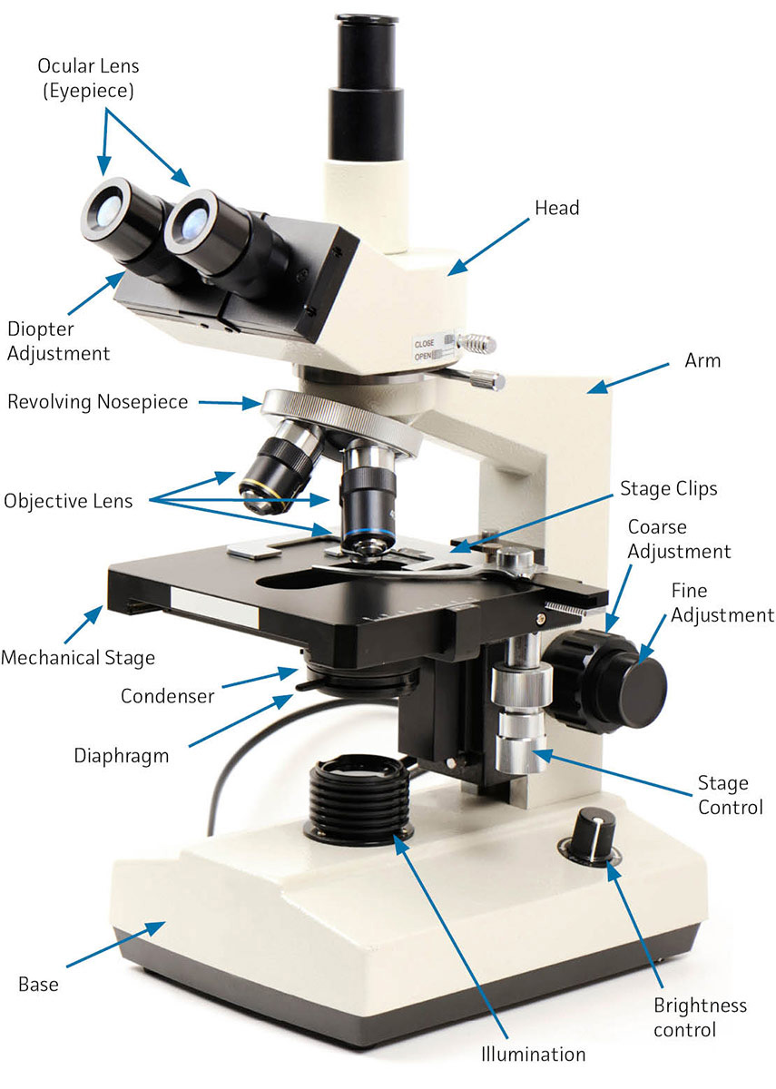
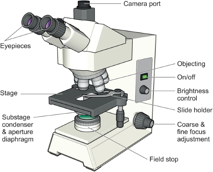
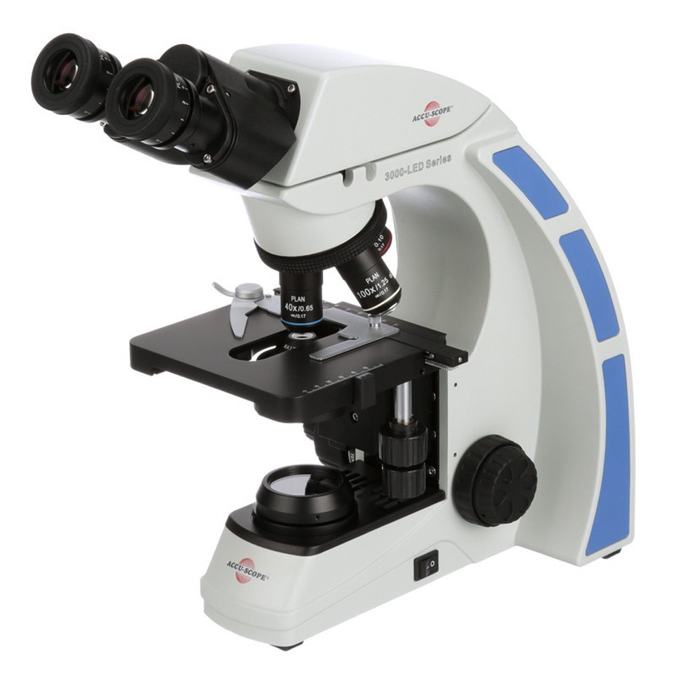
38 label the image of a compound light microscope using the terms provided
International News & Views | Reuters United Kingdom UK's Truss pledges to make the tough decisions as she sets out pitch to be PM, article with image July 13, 2022 Macro Matters UK house price growth falls to lowest since March 2021 ... Microscopy Lab Quiz Flashcards | Quizlet Label the image of a compound light microscope using the terms provided. If leaving an objective lens over the stage when storing the microscope, which objective lens should be placed over the stage? A. High power 40X B. Scanning 4X C. Oil immersion 100X D. Low power 10X B. Scanning 4X The fine adjustment knob on the microscope
A & P Microscope and Cell cycle Flashcards - Quizlet Label the image of a compound light microscope using the terms provided. Left side -rotating nosepiece -objective lenses -slide holder finger -stage -iris diaphragm lever -condenser -substage illuminator (lamp) -mechanical stage (control knob) Right side -eyepiece -light switch -course adjustment knob -fine adjustment knob

Label the image of a compound light microscope using the terms provided
PDF The Compound Light Microscope lab - Caldwell-West Caldwell Public Schools Part B: Care and handling of a compound light microscope Procedure: Answer the following questions concerning the care and handling of a compound light microscope. 1. Why is it important to carry the microscope correctly? 2. Why does the microscope need to be set at least 5cm away from the edge of the table? 3. en.wikipedia.org › wiki › Optical_microscopeOptical microscope - Wikipedia The optical microscope, also referred to as a light microscope, is a type of microscope that commonly uses visible light and a system of lenses to generate magnified images of small objects. Optical microscopes are the oldest design of microscope and were possibly invented in their present compound form in the 17th century. Label the parts of the light microscope below using provided reference ... 4. compound light, transmission electron, light electron, scanning electron . 5. The ability of a microscope to show details clearly is called 6. The ability of a microscope to increase an object's apparent sized is called . 7. To view a specimen with a light microscope, the specimen must be sliced _or be very small Course Section 1.
Label the image of a compound light microscope using the terms provided. Label the image of a compound light microscope using the terms provided. Exercise 1A -Parts of the compound microscope Write the correct label for each part of the microscope shown below: Exercise 1B - Using the compound microscope Match each part of the compound microscope on the left with its function on the right base... Posted 2 months ago View Answer 1. Microscope parts/labeling 9 Label the image of a compound light ... Microscope parts/labeling 9 Label the image of a compound light microscope using the terms provided. 1 points eyepiece eyepiece light source References References base arm slide holder arm stage mechanical stage fine adjustment knob power switch objectives Mindeneer Microscopy Lab Homework Saved... achieverstudent.comAchiever Student: The best way to upload files is by using the “additional materials” box. Drop all the files you want your writer to use in processing your order. If you forget to attach the files when filling the order form, you can upload them by clicking on the “files” button on your personal order page. Compound Microscope - Diagram (Parts labelled), Principle and Uses Also called as binocular microscope or compound light microscope, it is a remarkable magnification tool that employs a combination of lenses to magnify the image of a sample that is not visible to the naked eye. Compound microscopes find most use in cases where the magnification required is of the higher order (40 - 1000x).
› homework-help › questions-andSolved Label the image of a compound light microscope using ... Step-by-step answer. Who are the experts? Experts are tested by Chegg as specialists in their subject area. We review their content and use your feedback to keep the quality high. Transcribed image text: Label the image of a compound light microscope using the terms provided. BIO 168 Module 2 Quiz Review Flashcards | Quizlet Each of the following steps are necessary in preparing and observing a wet mount. Place the steps in the correct order. 1. Obtain a clean slide and cover slip. 2. Using a transfer pipette, obtain a drop of specimen and place onto the center of the slide. Parts of a microscope with functions and labeled diagram Q. List down the 18 parts of a Microscope. 1. Ocular Lens (Eye Piece) 2. Diopter Adjustment 3. Head 4. Nose Piece 5. Objective Lens 6. Arm (Carrying Handle) 7. Mechanical Stage 8. Stage Clip 9. Aperture 10. Diaphragm 11. Condenser 12. Coarse Adjustment 13. Fine Adjustment 14. Illuminator (Light Source) 15. Stage Controls 16. Base 17. 11 . O 2. Label the parts of the compound light micr... - Biology - Kunduz Names of these parts and. 11 . O 2. Label the parts of the compound light microscope on the diagram provided below (Figure 2-3). Names of these parts and their functions must be known to use the microscope correctly. ocular lens: remagnifies the image formed by the objective lens body tube: holds the lens system of the instrument arm: connects ...
di.uq.edu.au › files › 3522Introduction to Cell & Molecular Biology Techniques ... Fluorescence occurs when light of one wavelength “excites” a material and causes it to emit light of a different wavelength. Most fluorescent materials give off visible light after excitation by ultraviolet light. Structures may be naturally fluorescent (autofluorescence) or they may be labeled with a compound which is fluorescent (eg. Virtual Lab 2 Microscopy and Cells_for Hybrid absense.docx... Virtual Lab 2 - Microscopy and Cells BIO101 General Biology I ACTIVITY 1: MICROSCOPY Read the Introduction on page 23 of the lab manual. The Dissecting Microscope (aka: stereoscope): Watch the following YouTube video on the parts, function, and usage of the Leica EZ4 dissecting microscope (which is the model we have in the BIO101 lab rooms): o Page 23, bottom: Label the following image of a ... www1.udel.edu › biology › ketchamMicroscopy Pre-lab Activities - University of Delaware Microscope controls: turn knobs (click and hold on upper or lower portion of knob) throw switches (click and drag) turn dials (click and drag) move levers (click and drag) changes lenses (click and drag on objective housing) select a specimen (click on a slide) recorder.butlercountyohio.org › search_records › subdivisionWelcome to Butler County Recorders Office Copy and paste this code into your website. Your Link Name
Label the image to review the components of a compou... - Biology Finally, you will be given some follow-up concept check questions to make sure you understand the concept fully. 1. Both human and bacterial cells divide by mitosis. [Click to select) 2 Cells must their DNA prior to cell division replicate hydrolyze transcribe translate 3. Environmental factors control microbial growth through their Influence ...
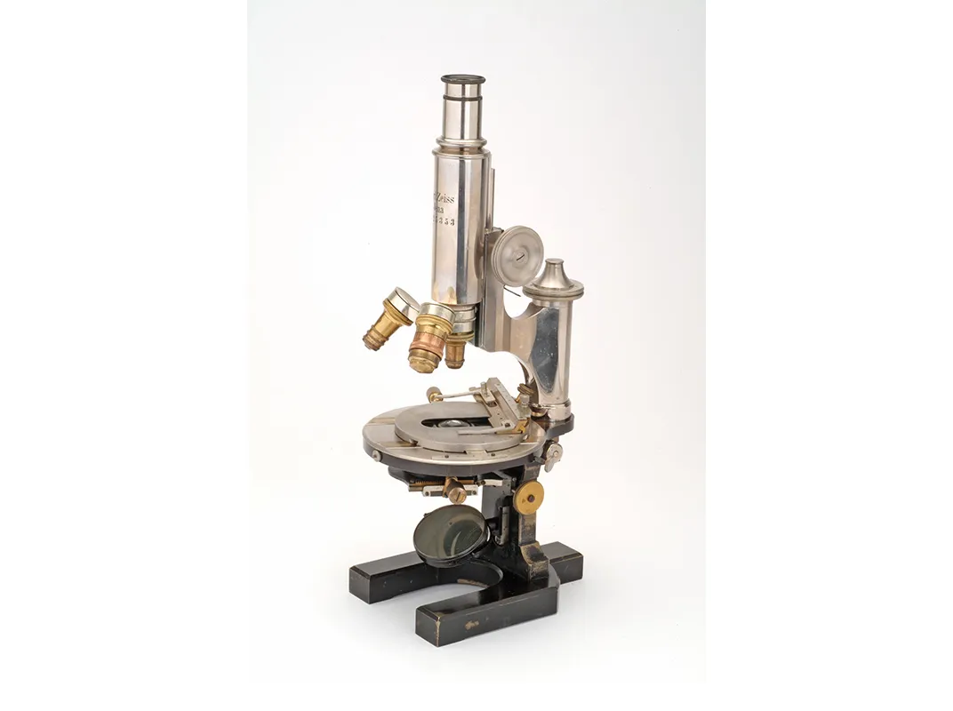
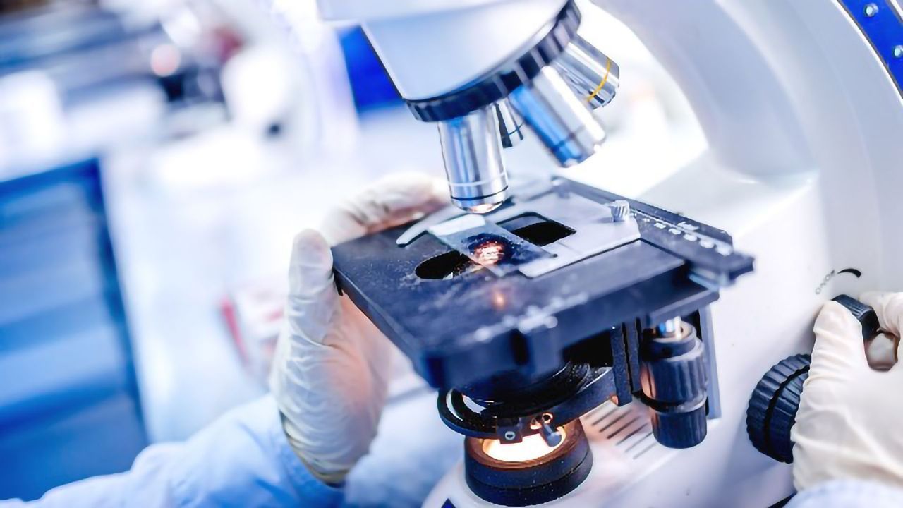
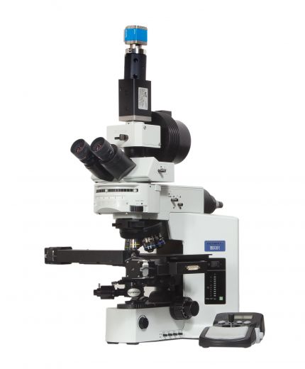
Compound Microscope: Definition, Diagram, Parts, Uses, Working ... - BYJUS A compound microscope is defined as. A microscope with a high resolution and uses two sets of lenses providing a 2-dimensional image of the sample. The term compound refers to the usage of more than one lens in the microscope. Also, the compound microscope is one of the types of optical microscopes. The other type of optical microscope is a ...
Solved Label the image of a compound light microscope using - Chegg Question: Label the image of a compound light microscope using the terms provided. Iris diaphragm lever Eyepiece Light switch Objective lenses Fine adjustment knob Rotating noseplece Stage Slide holder finger Substage illuminator (amp) Mechanical stage control knob One More Course adjustment knob Reset Condenser This problem has been solved!
Parts of the Microscope with Labeling (also Free Printouts) Image 2: The body tube part of a microscope is where the ray of light is bent to allow the object being viewed to enlarge by the scope. Picture Source: slideplayer.com 3. Turret/Nose piece. It is the revolving part of the microscope. It allows the use of different types of objective lenses by simply rotating the top part of the turret.
2 using a compound light microscope observe each Obtain a prepared root nodule slide and view under the compound light microscope. b. Compare the specimen to the image in your atlas. c. Draw a picture in the space provided on page 10 and identify the following: bacteroids (cells infected with Rhizobium), root hairs (if present). Indicate total magnification. Part 3.
compound microscope parts (labeling) Flashcards | Quizlet Start studying compound microscope parts (labeling). Learn vocabulary, terms, and more with flashcards, games, and other study tools. ... light source of the microscope. what is 8? eyepiece (ocular lens) - magnifying piece that is looked into in order to see the specimen ... knob that brings the image to a sharper focus. what is 13? base - the ...
Labeling the Parts of the Microscope Labeling the Parts of the Microscope This activity has been designed for use in homes and schools. Each microscope layout (both blank and the version with answers) are available as PDF downloads. You can view a more in-depth review of each part of the microscope here. Download the Label the Parts of the Microscope PDF printable version here.
Microscope parts/labeling Label the image of a compound light ... 1. State a procedures which should be used to properly handle a light microscope. 2. Explain why the light microscope is also called the compound microscope. 3- Images observed under the light microscope are reversed and inverted.
Label the image of a compound light microscope using the terms provided. Microscope parts/labeling 9 Label the image of a compound light microscope using the terms provided. 1 points eyepiece eyepiece light source References References base arm slide holder arm stage mechanical stage fine adjustment knob power switch... Posted 17 days ago Q: 1.
Bio 232 ~ Lab Midterm Flashcards - Quizlet Terms in this set (192) Label the body cavities in the figure. Identify the organ system based on the image and hint that provides the function of the system. Not all labels will be used. Place each of the labels in the box designating which plane or section is being referred to or demonstrated.
Compound Microscope Parts - Labeled Diagram and their Functions - Rs ... The term "compound" refers to the microscope having more than one lens. Basically, compound microscopes generate magnified images through an aligned pair of the objective lens and the ocular lens. In contrast, "simple microscopes" have only one convex lens and function more like glass magnifiers.
Label the above components of the compound light microscope A Ocular ... Keiser University Online Microbiology I Virtual Lab 1 Worksheet Microscopy & Gram Staining microscope b. resolution The power to show details clearly. The optic ability to distinguish detail such as the separation of closely adjacent objects. c. contrast converts phase shifts in light passing through a transparent specimen to brightness changes in the image
PDF The Compound Light Microscope The Compound Light Microscope TASK Refer to page 605 in your text to: 1. Name each of the structures described in the table to the right. 2. Match each structure to the letter in the diagram below. ** ALWAYS USE TWO HANDS TO CARRY A MICROSCOPE** Letter Structure Function joins body tube to base supports the entire microscope
Labelled Diagram of Compound Microscope - Biology Discussion The below mentioned article provides a labelled diagram of compound microscope. Part # 1. The Stand: The stand is made up of a heavy foot which carries a curved inclinable limb or arm bearing the body tube. The foot is generally horse shoe-shaped structure (Fig. 2) which rests on table top or any other surface on which the microscope in kept.
Label the parts of the light microscope below using provided reference ... 4. compound light, transmission electron, light electron, scanning electron . 5. The ability of a microscope to show details clearly is called 6. The ability of a microscope to increase an object's apparent sized is called . 7. To view a specimen with a light microscope, the specimen must be sliced _or be very small Course Section 1.
en.wikipedia.org › wiki › Optical_microscopeOptical microscope - Wikipedia The optical microscope, also referred to as a light microscope, is a type of microscope that commonly uses visible light and a system of lenses to generate magnified images of small objects. Optical microscopes are the oldest design of microscope and were possibly invented in their present compound form in the 17th century.
PDF The Compound Light Microscope lab - Caldwell-West Caldwell Public Schools Part B: Care and handling of a compound light microscope Procedure: Answer the following questions concerning the care and handling of a compound light microscope. 1. Why is it important to carry the microscope correctly? 2. Why does the microscope need to be set at least 5cm away from the edge of the table? 3.

.jpg)















:max_bytes(150000):strip_icc()/GettyImages-1146051036-66fd96c9f55a4d418422afeea05bfc9d.jpg)
Post a Comment for "38 label the image of a compound light microscope using the terms provided"