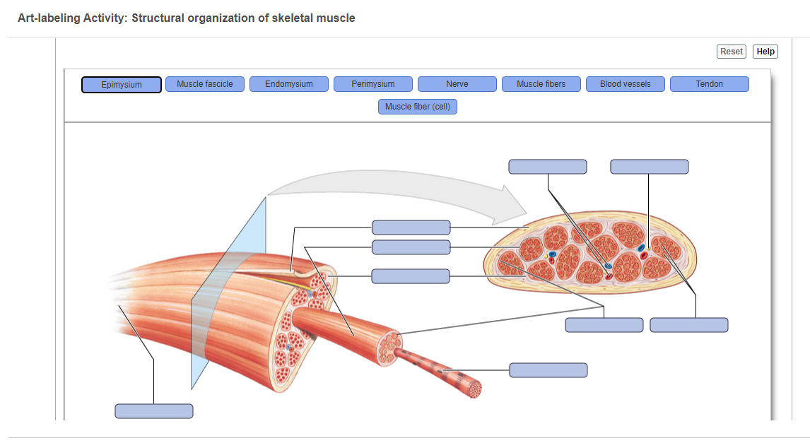
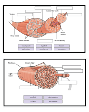
41 art-labeling activity: structure of muscle tissues
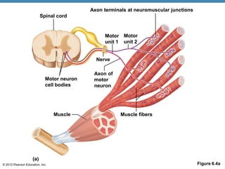
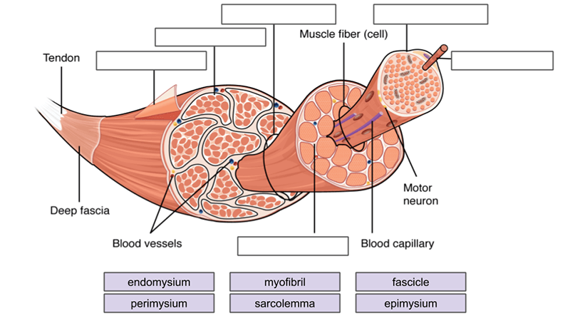
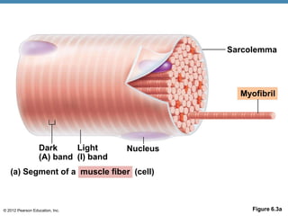
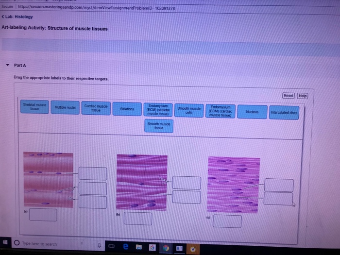
Art-labeling Activity: The Structure of a Skeletal Muscle Fiber Start studying Art-labeling Activity: The Structure of a Skeletal Muscle Fiber. Learn vocabulary, terms, and more with flashcards, games, and other study tools. ... Write. Test. PLAY. Match. Created by. BabeRuthless0504. Terms in this set (2) Art-labeling Activity: The Structure of a Skeletal Muscle Fiber... Art-labeling Activity: The Structure ... Solved Secure https:/ C Lab: Histology Art-labeling | Chegg.com Answer A is skeletal mucle tissues consisting of = ENDO …. View the full answer. Transcribed image text: Secure https:/ C Lab: Histology Art-labeling Activity: Structure of muscle tissues 102091378 Part A Drag the appropriate labels to their respective targets tissue Smooth muSECM) (cardiac tissue (ECM) (skeletal Nucleus issue Type here to ...
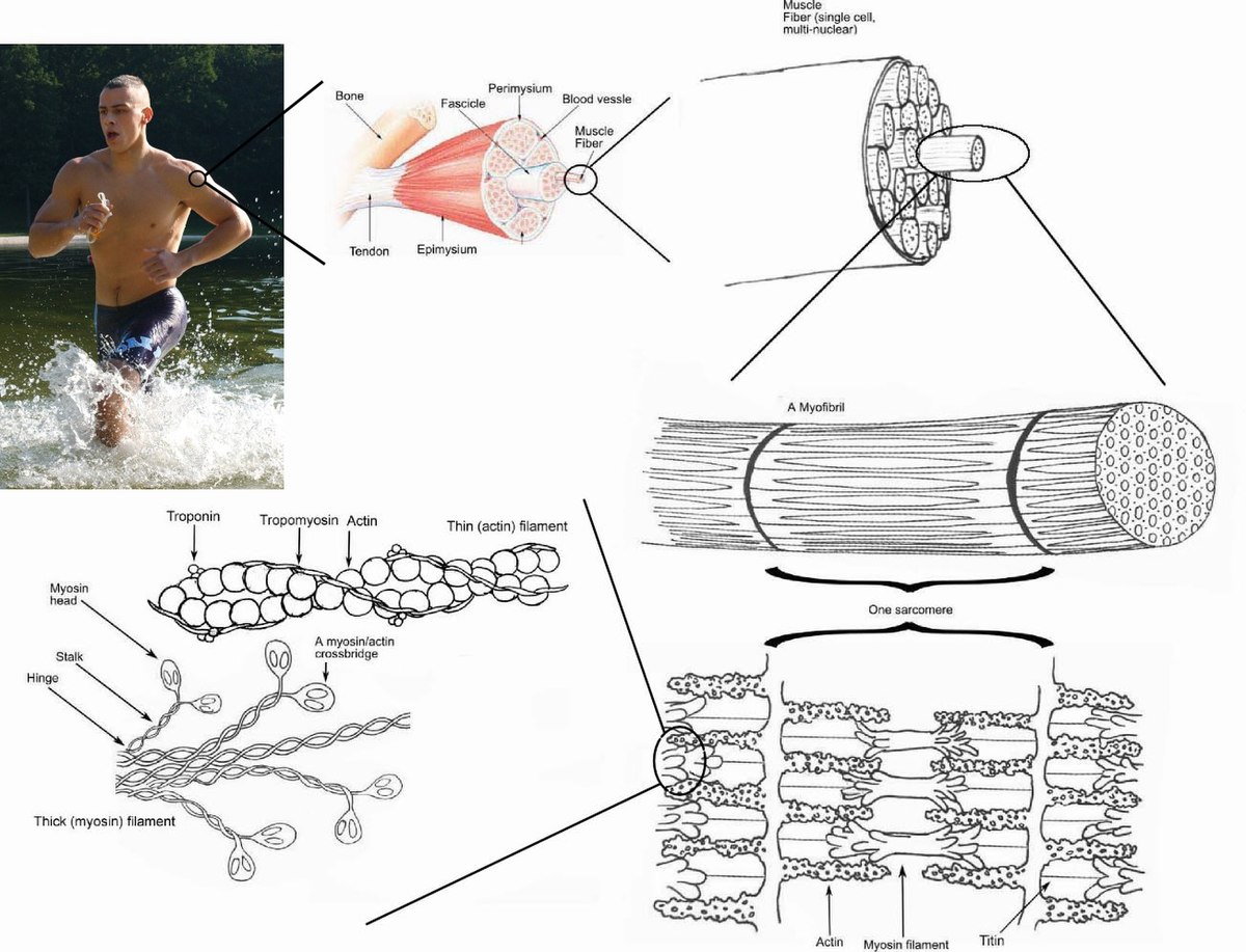
Answered: Art-labeling Activity: Structural… | bartleby Q: Label different areas of an individual muscle unit known as a sarcomere below: Actin A Band Mine… A: The smallest functional unit of muscle tissue is called sarcomere. It consist of actin and myosin…
Art-labeling activity: structure of muscle tissues
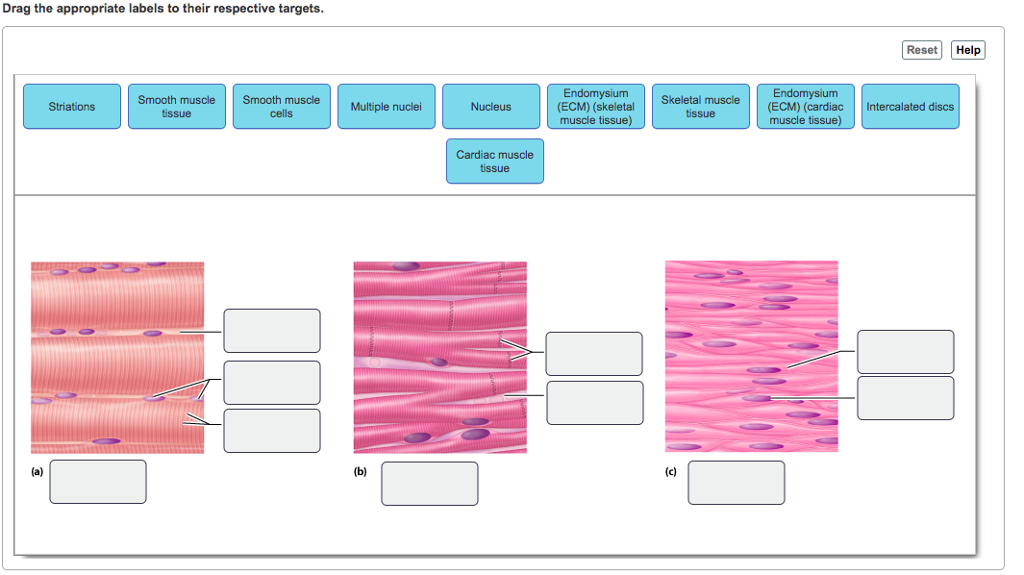
Solved Art-labeling Activity: Histology of Muscle Tissue 15 - Chegg Art-labeling Activity: Histology of Muscle Tissue 15 of 22 Reset Help intercalated discs Skelet muscle sbor Cardiac muscle cells Nucleus smooth muscle Smooth muscle cos Strations cardiac musco) Strabons skola muscel Nucle (candid muscle) Cardia Smoom muscle tissue muscle tissue muice tissu Nucle skoletai musco) art-labeling activity: the structure of a sarcomere Solved Art Labeling Activity The Structure Of A Sarcomere Chegg Com Organ Systems and Body Cavities From Allen and Harper Laboratory Manual for Anatomy and Physiology 5 th Edition 21 T he cells. Anatomy and Physiologyb 2 ml of water was added to the control test tubeTherefore it is not enough to be able to identify a structure its function must ... Art-labeling Activity: Structure of a Typical Synovial Joint Art labeling activity -structure of a nail superficial and cross-sectional viewsjpg. Sets found in the same folder. Structure of a nail. Fibrous Capsule outer- provides joint stability 2. The mandible is attached to the rest of the skull by a freely movable joint. Curves and Regions of the Vertebral Column Learning Goal.
Art-labeling activity: structure of muscle tissues. Art labeling Activity The Structure of a Skeletal Muscle Fiber Drag the ... Art labeling Activity The Structure of a Skeletal Muscle Fiber Drag the labels from AA 1. Study Resources. Main Menu; by School; by Literature Title; by Subject; ... Art labeling activity the structure of a skeletal. School No School; Course Title AA 1; Uploaded By JusticeWillpower7289. Pages 80 Answered: Art-labeling Activity: Structural… | bartleby Transcribed Image Text: Art Labeling Activity the Structure of the Epidermis Summary of epithelial tissues Part A Drag the appropriate labels to their respective targets. Part A Diagram of. The structure of the epidermis. Lab - Integumentary System 226 Correct Art-Labeling Activity. Part A Drag the labels onto the epidermal layers. What effect would you see in the most superficial epidermal layers. Art-labeling Activity: Structure of a Typical Synovial Joint Art labeling activity -structure of a nail superficial and cross-sectional viewsjpg. The elbow is the joint connecting the upper arm to the forearm. Types of Synovial Joints. In synovial joints the ends of. Label the diagram of a typical synovial joint using the terms provided in. Muscles of the Chest Abdomen and Thigh Deep Dissection 1 of 2jpg.
EOF Art-labeling Activity: Types of Connective Tissue Proper Start studying Art-labeling Activity: Types of Connective Tissue Proper. Learn vocabulary, terms, and more with flashcards, games, and other study tools. chapter 9- Mastering A and P, Chapter 9-1 The muscular Tissue Art-labeling Activity: The structure of a skeletal muscle fiber. PICTURE. Chapter Test - Chapter 9 Question 3 ... Art-labeling Activity: Phases and significant events of a muscle twitch in the myogram. PICTURE. ... The muscle tissue in the meat would probably not become stiff after death if it still had enough a) Calcium ... art-labeling activity: structure of the epidermis The structure indicated by label E is part of which of the following. Basal cells in the stratum germinativum divide adding in new daughter cells and pushing the cells above upward through the layers. A finger-like projection or fold known as the dermal papilla. To supply cells to replace those lost.
BIO 200 Chapter 9 - Muscle Tissue Physiology Flashcards - Quizlet The storage and release of calcium ions is the key function of the: sarcoplasmic reticulum. A group of skeletal muscle fibers together with the surrounding perimysium form a (n): fascicle. Art-Ranking Activity: Stages of an action potential. A crossbridge forms when: a myosin head binds to actin. art-labeling activity: sarcomere structure - leticiavandeputte A sarcomere is the area between two Z lines that can be regarded the fundamental structural and functional unit of muscle tissue. The thick filaments in the A band and thin filaments in the I band interact and. Start studying Art-labeling Activity. 42322 503 PM Week 3 Chapter 9 810 Correct Art-labeling Activity. art-labeling activity: structure of a long bone View art labeling activity - the vertebral columnjpg from ANT MISC at Miami Dade College Miami. Mastering A P Chapter 6 Bones And Skeletal Tissues Flashcards Quizlet Part A Drag the labels to identify the structures of a long bone.. With high mitotic activity and they are the only bone cells that divide. Label the types of bone cells. A long ... Answer correct art based question chapter 4 question - Course Hero ANSWER: Correctmultinucleate cells branched cells intercalated discs situated between cells striations tendons and ligaments attached to bones heart ducts of certain glands dense irregular connective tissue smooth muscle tissue skeletal muscle tissue cardiac muscle tissue
[Solved] Art-Labeling Activity: | Course Hero Composed of actively dividing keratinocytes with spinous-like projections (prickle cells) This layer produces keratin and induces keratinization. Langerhans cells are also located in this layer. Stratum basale (also called the basal cell layer of the epidermis)
A&P 1- CHAPTER 9 MASTERING ASSIGNMENTS Flashcards - Quizlet Art-labeling Activity: The structure of a skeletal muscle fiber PICTURE Which thin filament-associated protein binds two calcium ions? troponin Action potential propagation in a skeletal muscle fiber ceases when acetylcholine is removed from the synaptic cleft.
Art-labeling Activity: Structure of a Typical Synovial Joint Art labeling activity -structure of a nail superficial and cross-sectional viewsjpg. Sets found in the same folder. Structure of a nail. Fibrous Capsule outer- provides joint stability 2. The mandible is attached to the rest of the skull by a freely movable joint. Curves and Regions of the Vertebral Column Learning Goal.
art-labeling activity: the structure of a sarcomere Solved Art Labeling Activity The Structure Of A Sarcomere Chegg Com Organ Systems and Body Cavities From Allen and Harper Laboratory Manual for Anatomy and Physiology 5 th Edition 21 T he cells. Anatomy and Physiologyb 2 ml of water was added to the control test tubeTherefore it is not enough to be able to identify a structure its function must ...
Solved Art-labeling Activity: Histology of Muscle Tissue 15 - Chegg Art-labeling Activity: Histology of Muscle Tissue 15 of 22 Reset Help intercalated discs Skelet muscle sbor Cardiac muscle cells Nucleus smooth muscle Smooth muscle cos Strations cardiac musco) Strabons skola muscel Nucle (candid muscle) Cardia Smoom muscle tissue muscle tissue muice tissu Nucle skoletai musco)






:watermark(/images/watermark_only_sm.png,0,0,0):watermark(/images/logo_url_sm.png,-10,-10,0):format(jpeg)/images/anatomy_term/skeletal-muscle/0kdrKFzTosiUmeSgQIFJjQ_Skeletal_muscle_01.png)









Post a Comment for "41 art-labeling activity: structure of muscle tissues"