43 sheep eye diagram labeled
PDF PRACTICE SHEEP PARTS - Washington State University PRACTICE SHEEP PARTS : ... Back Thigh Eye Dock Dewclaw Ear Belly Neck Mouth Face Ribs Knee Loin Poll Stifle Foreflank Hoof Rearflank Pastern Forearm Shank ... Next to each part below write in the NUMBER for the part on the hog diagram. Sheep Project - Practice Parts : Junior / Intermediate / Senior (Circle One) NAME . ANSWER KEY : CLUB . internal eye anatomy sheep brain labeled anatomy external lobe dissection frontal physiology nervous system occipital spinal cord savalli Cow Anatomy Diagram Showing Internal Organs | Vintage Antique Ephemera organs anatomy cattle Orbit & Eye - Atlas Of Anatomy doctorlib.info
PDF Sheep Eye Dissection - mrgscience.com Place the sheep's eye on it for inspection. 2. Use a pencil to sketch a side-view external diagram of the eyeball in Observation #1. Make sure to label the sclera, cornea, optic nerve, and muscles. 3. Cut the fat and muscles off the eyeball so that you can see the sclera. 4. Place your eye specimen in the dissection pan.

Sheep eye diagram labeled
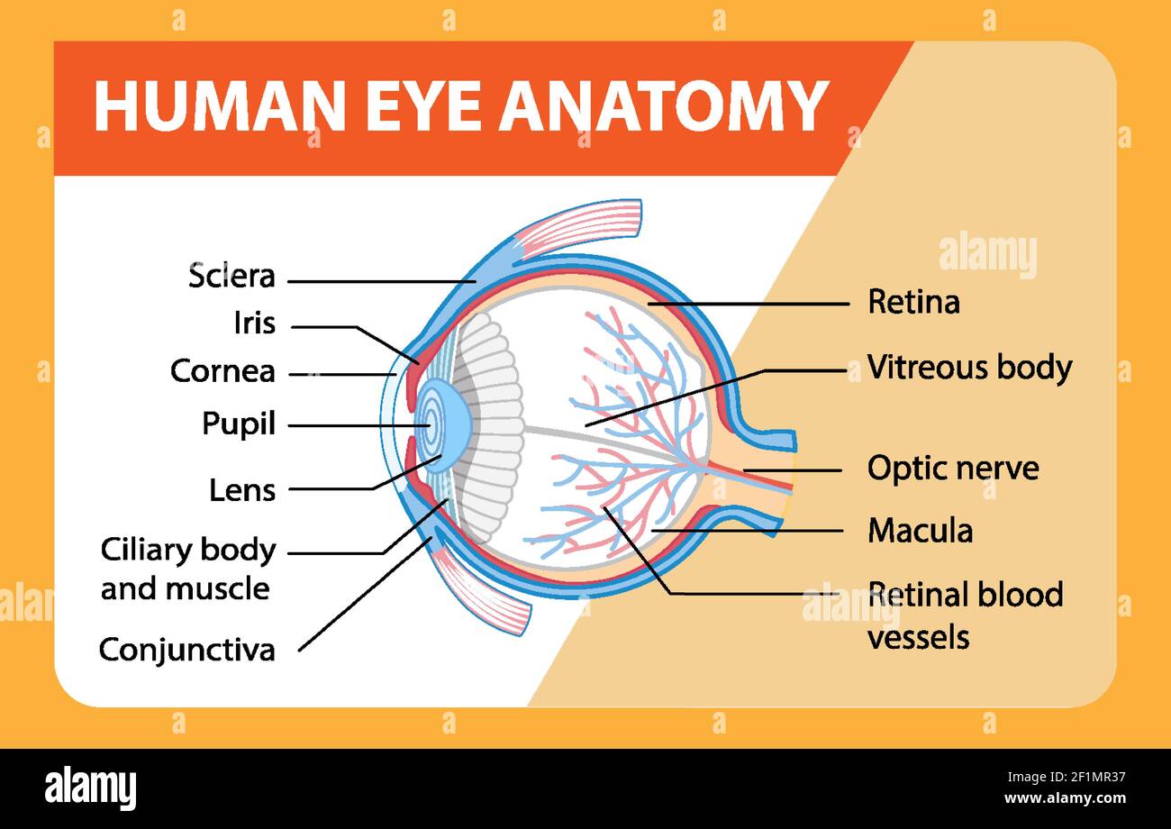
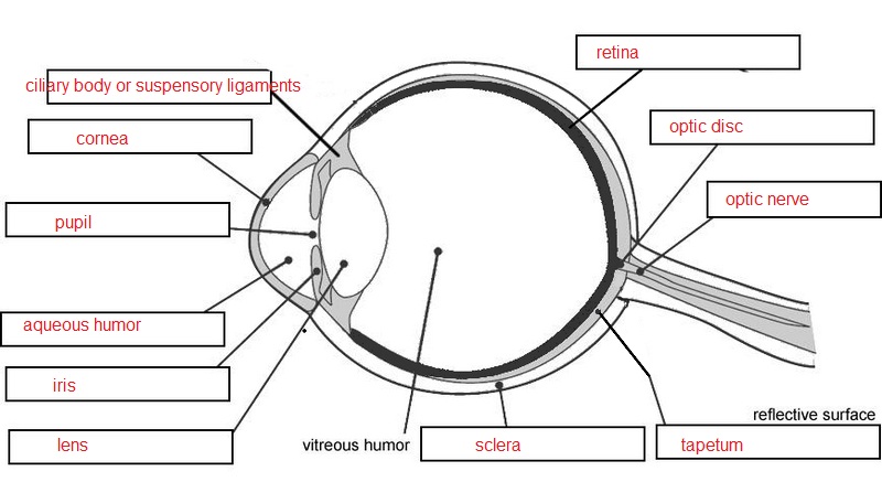
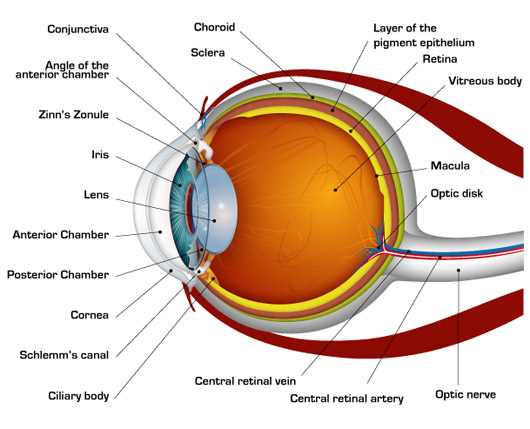
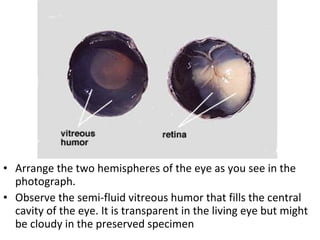
Eye Anatomy - John Burroughs School 1. Print a Diagram of the Eye - Click on this link and then use the browser print command to produce a diagram to use with the student tutorial. 2. Label the eye diagram. 3. Identify the parts of the eye (model). 4. Virtual sheep eye dissection. 5. Test your knowledge of the sheep eye. Bruce Westling, Photographer and Web Page Author PDF Dissecting and Diagramming the Eye - Environmental Science Students should create a rough draft of their eye diagrams (viewed from different angles, such as a top view and a side view and a frontal view). Parts to label could include sclera, cornea, lens, vitreous body, iris, pupil, retina, and optic nerve. They may trade drafts with a partner to analyze if any parts or details might be missing. DOC Sheep Eye Dissection - PC\|MAC Move the retina to see the dark, metallic-looking tissue at the back of the eye. This is the choroid. The portion that appears iridescent blue and green with shades of yellow is called the tapetum. Assignment: Use the following glossary to label the eye diagram below. Aqueous humor: clear fluid filling the area between the lens and cornea ...
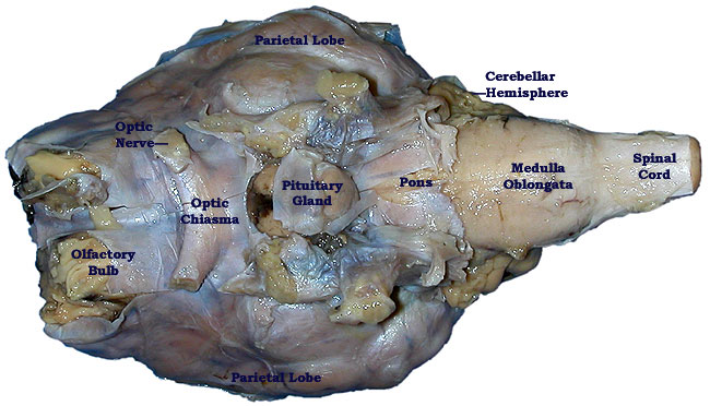
Sheep eye diagram labeled. diagram of a cow eye sheep eye dissection worksheet LAS Assignment 7 - Eye Enucleation cmapsconverted.ihmc.us eye cow labeled cows enucleation unlabeled coronal section bio201 return preserved Sheep eye dissection worksheet. Sheep brain dissection lab ventral inferior anatomy label human bi diagrams companion locate following biologyjunction. sheep internal anatomy eye cow dissection anatomy labeled diagram sheep eyes psychology homepages wmich edu department biology human eyeball dissected parts cat. BIO201-Sheep Brain httwww.savalli.us. bio201 savalli unlabeled. Sheep Heart Open 1 Answers . anemic infarct vein. Sheep External Anatomy - PurposeGames . purposegames. Sheep ... anatomy of sheep eye eye anatomy cow human dissection lab nervous structure labeled diagram parts anterior tunic spinal cord system internal external biology physical Heart Anatomy - Vessels heart vessels sheep labeled anatomy pulmonary right virtual thicker Sheep brain dissection. Smith, k / anatomy and physiology activities. Sheep Brain Dissection with Labeled Images 1. The sheep brain is enclosed in a tough outer covering called the dura mater. You can still see some structures on the brain before you remove the dura mater. Take special note of the pituitary gland and the optic chiasma. These two structures will likely be pulled off when you remove the dura mater. Brain with Dura Mater Intact
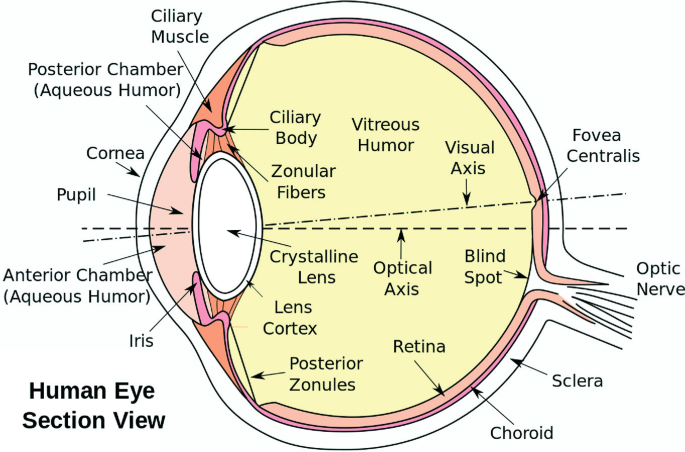
Cow Eye Dissection Teaching Resources | Teachers Pay Teachers This is a comprehensive dissection guide of the cow eye, designed for a high school or early college Biology or Anatomy & Physiology class. The guide includes step-by-step instructions and labeled diagrams that will lead students through the external anatomy of the eye, followed by dissection of the internal structures. Diagram of Eye Anatomy Biology: Sheep Eye Dissection albertsscientist4com.blogspot.com. eye sheep dissection external cornea dissected optic nerve structures biology left. Astronomy diagrams concept resources orbit points universe planets inferior starry night starrynighteducation. Biology: sheep eye dissection. Starry night education PDF Part I: The Eye - OKALOOSA SCHOOLS Draw the eye diagram to the right and label the following structures: 1. Lens 2. Cornea 3. Aqueous humor 4. Pupil 5. Iris 6. Ciliary Muscle 7. Sclera 8. Vitreous Humor 9. Retina 10. Blind Spot 11. Choroid Layer 12. Optic Nerve 13. Tapetum Lucidum Cornea Cornea-transparent membrane that covers iris and pupil. Focuses light on the retina. diagram of the eye labeled Human Eye: Anatomy, Parts And Structure . eye anatomy structure human parts. Sheep Brain External View Labeled | Brain Anatomy, Occipital Lobe . sheep brain labeled anatomy external lobe dissection frontal physiology nervous system occipital spinal cord savalli. Topic: Flatworms ...
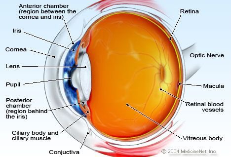
Heart Diagram Labeled Igcse : The Human Eye Edexcel Igcse Biology ... This diagram shows the parts of the eye. …atrial septum may involve the atrioventricular valves and may be associated with incompetence of these valves. Sclera tapetum optic nerve blind spot lens vitreous humor iris cornea retina aqueous humor pupil. Your left is the heart's right, which is why the lefts and rights are swapped in the diagram. human eye diagram Diagram of the eye. Neck, nerves of. Sheep brain labeled anatomy external lobe dissection frontal physiology nervous system occipital spinal cord savalli. Human Anatomy in Full Color | Scribd we have 9 Images about Human Anatomy in Full Color | Scribd like Human Eye: Anatomy, parts and structure, Diagram of the Eye | ClipArt ETC and also ... sheep eye anatomy baby rams lambs ram cute found animal sheep lamb too head same. NEW - Cow Eye Dissection: Video Lab Series - YouTube . cow eye dissection labeled lab. Sulcus coronary heart cat anterior auricles sheep vascular runs filled fat under. Brain sheep caudate coronal nucleus section psychology homepages wmich edu. Sheep eye dissection Lab #12: Sheep Eye Dissection Diagram | Quizlet a ring of muscle tissue that forms the colored portion of the eye around the pupil and controls the size of the pupil opening. Pupil the adjustable opening in the center of the eye through which light enters. Choroid middle, vascular layer of the eye, between the retina and the sclera. Palpebrae another name for eyelids; protects eye.
Cow's Eye Dissection - Eye diagram - Exploratorium A muscle that controls how much light enters the eye. It is suspended between the A cow's iris is brown. many colors, including brown, blue, green, and gray. A clear fluid that helps the cornea keep its rounded shape. The pupil is the dark circle in the center of your iris. It's a hole that Your pupil is round.
sheep eye anatomy Eye anatomy cow dissection labeled section human parts sagittal lab sheep lens eyes quizlet internal student physiology animal choroid heart. The resolution of the human eye is 576 megapixels. Sheep pig eyes dissection organs preserved pack expand
brain parts labelled diagram brain parts labelled diagram. Brain shark labeled nervous cranial dogfish system anatomy spinal dissected cavity nerves organs primitive human non animals very sense cord. Sheep brain labeled. Eye anatomy cow dissection labeled sheep section parts human lab sagittal lens eyes physiology quizlet internal student animal system choroid. Smith, K ...
human eye parts diagram Sheep Eye Dissection - YouTube . sheep eye dissection worksheet. Illustration Human Eye Anatomy Stock Vector 141161560 - Shutterstock . anatomy. Human Eye Anatomy Classroom Diagram Educational Chart Poster 24x36 Inch . Human Eye Anatomy Stock Vector. Illustration Of Diagram - 37354087
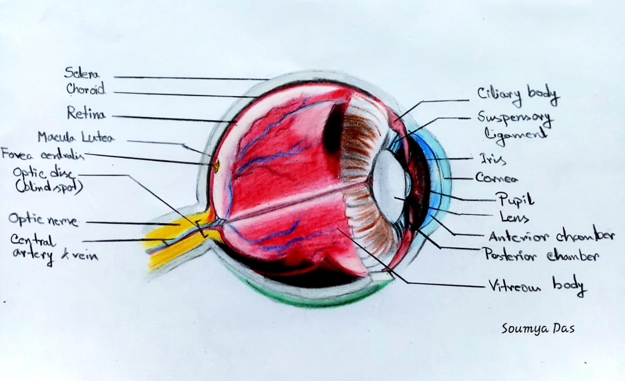
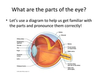
Sheep eye dissection - SlideShare Eye Dissection • Before we go over the dissection, let's review the parts of the eye and their function 12. Cross section You must know: 1. Lens 2. Cornea 3. Aqueous humor 4. Pupil 5. Iris 6. Ciliary Muscle 7. Sclera 8. Vitreous Humor 9. Retina 10. Blind Spot 11. Choroid Layer 12. Optic Nerve 13.
diagram of the eye labeled eye anatomy cow dissection labeled section human parts sagittal lab sheep lens eyes quizlet internal student physiology animal choroid heart. Chapter Two . brain sagittal human section anatomy labeled diagram cat robotspacebrain plane physiology drawing cut parts midsagittal study medical cranial diagrams system. Biology: Sheep ...
sheep anatomy diagram Diagram Of The Cow Eye Images How To Guide And Refrence | Cow Eyes, Eye . eye cow anatomy diagram labeled dissection eyeball eyes guide lucidum. Heart Dissection (Pig) - YouTube . heart dissection pig. Pin By Kenton Antiques On Vintage Antique Ephemera | Sheep, Ewe Sheep . sheep anatomy diagram ...
human eye diagram to label Sheep Eye Dissection - YouTube . sheep eye dissection. Pin On Examples Printable Label Templates . quizlet slidedocnow spinal cord. Stevens Anatomy: Basic Ear Worksheet stevensanatomy.blogspot.com. ear worksheet label diagram sound waves anatomy labeling key answer inner stevens incus worksheeto middle. Labeled ...
eye diagram labeled Eye sheep dissection external cornea dissected optic nerve structures biology left. Chapter two eye diagram labeled Chapter Two. 9 Pics about Chapter Two : Diagram of the Eye Quiz, Smith, K / Anatomy and Physiology Activities and also LAS Assignment 7 - Eye Enucleation. Chapter Two
Sheep Eye Diagram | Quizlet Sheep Eye Diagram | Quizlet Sheep Eye STUDY Learn Write Test PLAY Match Created by erm9606 PLUS Terms in this set (12) Cornea ... Anterior chamber ... Iris ... Pupil ... Lense ... Choroid ... Retina ... Vitreous body ... Sclera ... Macula ... Optic nerve ... Optic disk ... Sets found in the same folder Lobes of Brain 5 terms erm9606 PLUS Neuron
Practice Lab Practical on the Sheep Brain Identify the lobe labeled 1. Identify the lobe labeled 2. Identify the lobe labeled 3. Identify the structure labeled 4. Identify the structure lobe labeled 5. ... Identify the shiny membrane visible on the sheep brain surface. In the above picture: Identify the structure labeled 1. Identify the structure labeled 2. ...
DOC Sheep Eye Dissection - PC\|MAC Move the retina to see the dark, metallic-looking tissue at the back of the eye. This is the choroid. The portion that appears iridescent blue and green with shades of yellow is called the tapetum. Assignment: Use the following glossary to label the eye diagram below. Aqueous humor: clear fluid filling the area between the lens and cornea ...
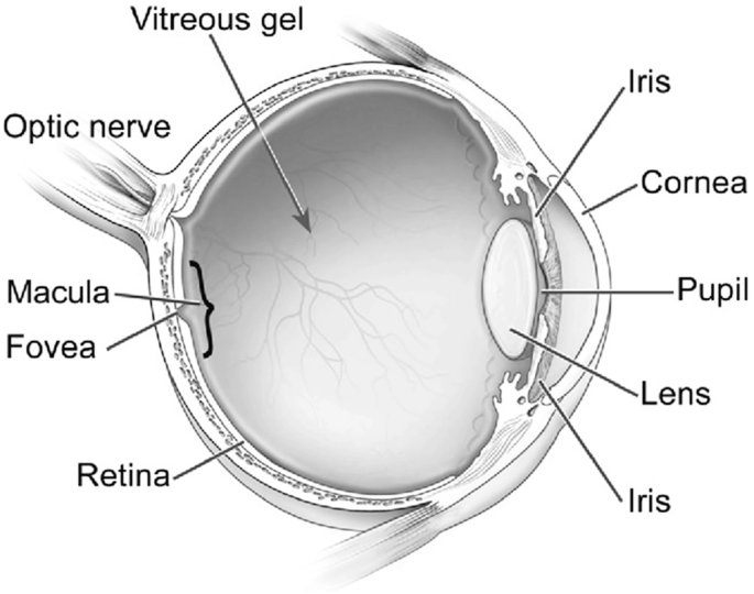
PDF Dissecting and Diagramming the Eye - Environmental Science Students should create a rough draft of their eye diagrams (viewed from different angles, such as a top view and a side view and a frontal view). Parts to label could include sclera, cornea, lens, vitreous body, iris, pupil, retina, and optic nerve. They may trade drafts with a partner to analyze if any parts or details might be missing.
Eye Anatomy - John Burroughs School 1. Print a Diagram of the Eye - Click on this link and then use the browser print command to produce a diagram to use with the student tutorial. 2. Label the eye diagram. 3. Identify the parts of the eye (model). 4. Virtual sheep eye dissection. 5. Test your knowledge of the sheep eye. Bruce Westling, Photographer and Web Page Author















Post a Comment for "43 sheep eye diagram labeled"