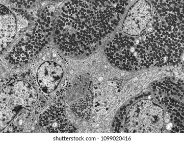
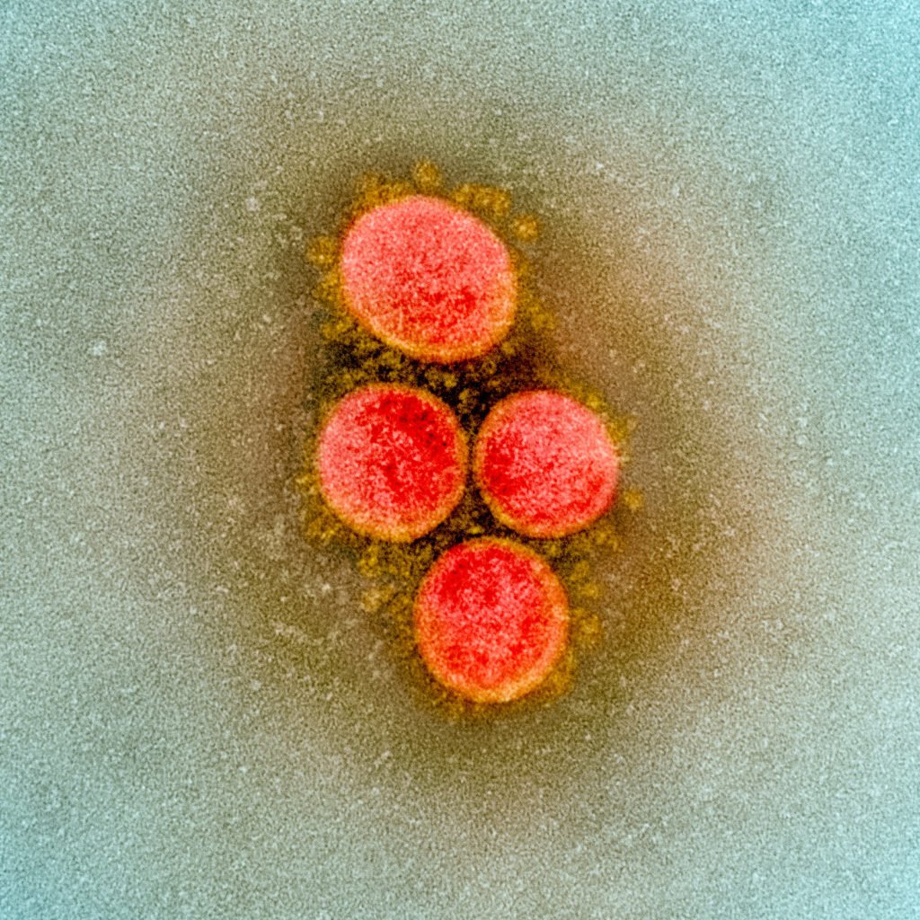
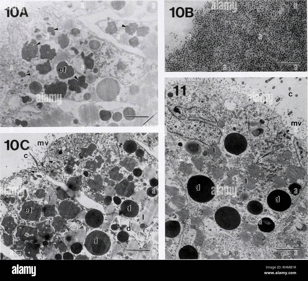
43 transmission electron micrograph labeled
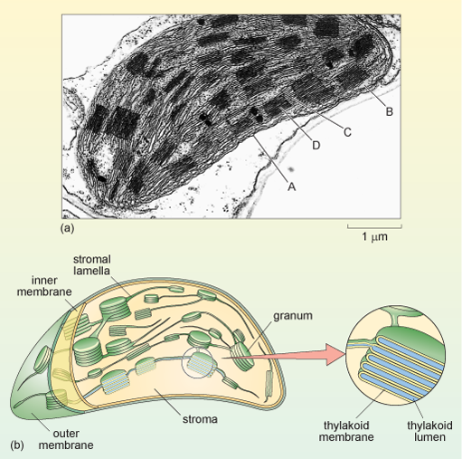
Correlative Fluorescence and Scanning Transmission Electron Microscopy ... Correlative fluorescence microscopy combined with scanning transmission electron microscopy (STEM) of cells fully immersed in liquid is a new methodology with many application areas. Proteins, in live cells immobilized on microchips, are labeled with fluorescent quantum dot nanoparticles. PDF Identifying Organelles from an Electron Micrograph The photograph shown below details chloroplast structure as viewed with a transmission electron microscope Courtesy of Dr. Julian Thorpe - EM & FACS Lab, Biological Sciences University Of Sussex A single Granum Chloroplast envelope visible as two membranes Stroma containing numerous small ribosomes Lamellae connecting different grana
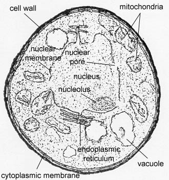
Solved Label the transmission electron micrograph of the | Chegg.com Transcribed image text: Label the transmission electron micrograph of the cell. 0 Nucleus rences Mitochondrion Heterochromatin Peroxisome Vesicle ULAR bumit Click and drag each label into the correct category to indicate whether it pertains to the cytoplasm or the plasma membrane.

Transmission electron micrograph labeled
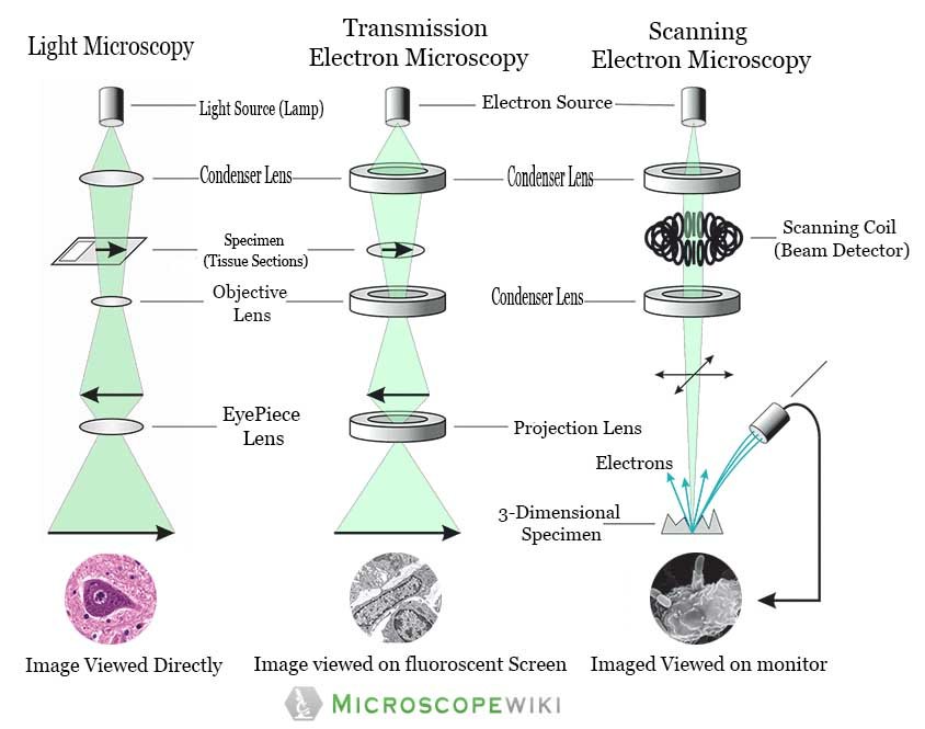
Transmission Electron Microscopy - an overview | ScienceDirect Topics Transmission electron microscopy (TEM) TEM was first described in 1932 by Knoll and Ruska, but the resolution of early models was no better than existing light microscopes. TEM resolution exceeded that of light microscopy within three years and, by 1946, resolution to ten Angstroms (1×10 −10 m) had been achieved. Transmission Electron Microscope (With Diagram) The final image in a TEM is known as transmission electron micrograph. The salts of some heavy metals, e.g., lead; osmium, tungsten and uranium are often used for staining. These heavy metal stains are used to increase the contrast between ultra structures and the background. Label This Transmission Electron Micrograph : TEM of chloroplast from ... Label this transmission electron micrograph of relaxed sarcomeres by clicking and dragging the labels to the correct location . Transmission electron microscopy (tem) is one of the oldest technologies and still. Molecular labeling for correlative microscopy: Fluorescence microscopy in combination with tem and an ion beam analysis (iba, which ...
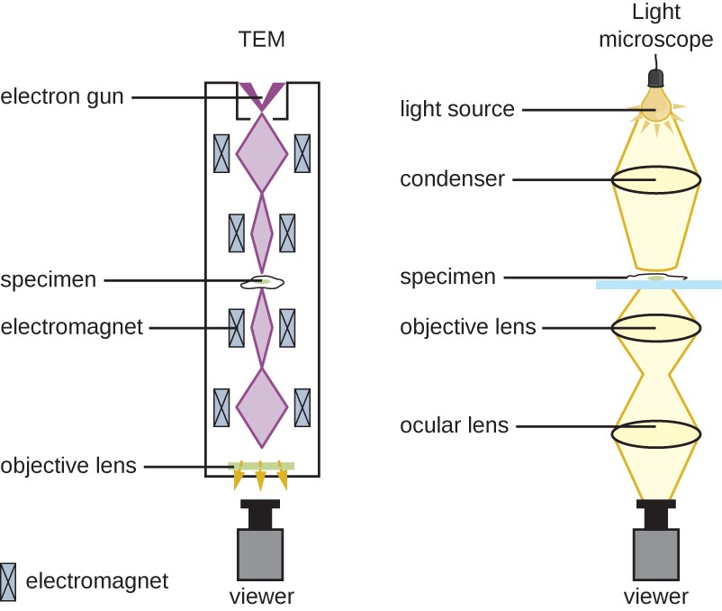
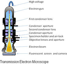
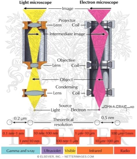
Transmission electron micrograph labeled. Microscope Types (with labeled diagrams) and Functions The shorter wavelength of electrons compared to visible light photons helps the observer achieve a very high resolving power compared to normal microscopes thereby aiding observers to see very tiny objects clearly. Electron microscope labeled diagram The different types of electron microscopes are: Transmission Electron Microscope Transmission Electron Microscopy - Penn State College of Medicine Research The lab is equipped with a high-resolution transmission electron microscopes (JEOL 1400) and a full suite of ancillary sample preparation equipment to fit most TEM-related needs. The equipment can provide nanometer-scale analysis for biological, chemical and materials science. The TEM facility offers multiple levels of service and training ... Solved Label the transmission electron micrograph of the | Chegg.com Experts are tested by Chegg as specialists in their subject area. We review their content and use your feedback to keep the quality high. Answer The label is indicated from TOP to BOTTOM Ciliu …. View the full answer. Transcribed image text: Label the transmission electron micrograph of the cilium. Microvillus Axoneme Cilium Dynein arm. Transmission electron microscopy characterization of ... - PubMed Transmission electron microscopy characterization of fluorescently labelled amyloid β 1-40 and α-synuclein aggregates Abstract Background: Fluorescent tags, including small organic molecules and fluorescent proteins, enable the localization of protein molecules in biomedical research experiments.
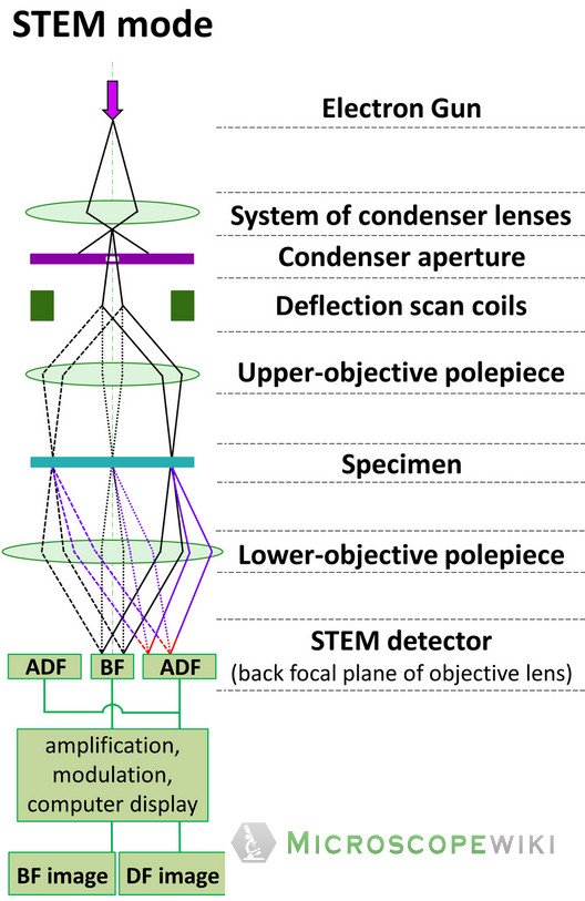
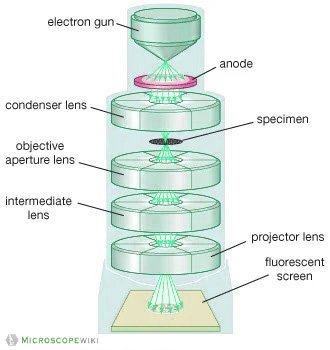
Transmission Electron Microscope (TEM)- Definition, Principle, Images Parts of Transmission Electron Microscope (TEM) Their working mechanism is enabled by the high-resolution power they produce which allows it to be used in a wide variety of fields. It has three working parts which include: Electron gun Image producing system Image recording system Electron gun Label This Transmission Electron Micrograph Of A Relaxed Sarcomere ... Label this transmission electron micrograph of relaxed sarcomeres by clicking and dragging the labels to the correct location . Label the following image using the terms provided. Note how the sarcomeres are extended to only approximately 120 % . IMG_2132 - FIGURES Label this transmission electron from Solved Label the transmission electron micrograph based on - Chegg Expert Answer nucleus is the house of the genetic material which contains all the h … View the full answer Transcribed image text: Label the transmission electron micrograph based on the hints provided Mitochondrion Heterochromatin Plasma cell Nucleus Rough endoplasmic reticulum Nucleolus Previous question Next question Electron Microscope-Definition, Principle, Types, Uses, Labeled Diagram The electron microscope is placed vertically and has the shape of a tall vacuum column. It consists of the following elements: 1. Electron gun. A heated tungsten filament that produces electrons makes up the electron cannon. 2. Electromagnetic lenses. The condenser lens directs the electron beam to the specimen.
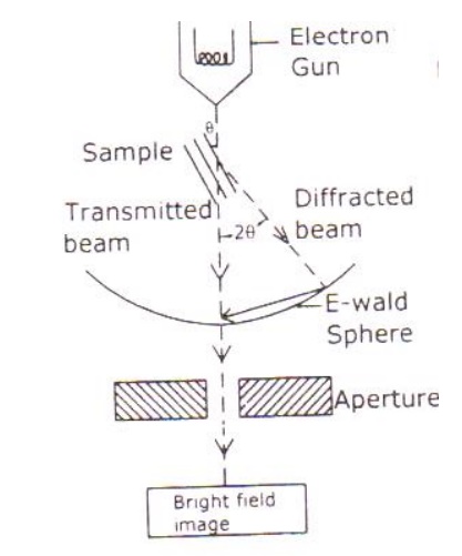
Transmission electron microscopy - Wikipedia Transmission electron microscopy ( TEM) is a microscopy technique in which a beam of electrons is transmitted through a specimen to form an image. The specimen is most often an ultrathin section less than 100 nm thick or a suspension on a grid. Transmission Electron Micrograph of transfected HL-1 cells labeled for ... Transmission Electron Micrograph of transfected HL-1 cells labeled for TMEM43 with immunogold. A and B. Single immunogold labeling experiments used 15 nm gold particles to label GFP. A.... Transmission electron microscopy DNA sequencing - Wikipedia Transmission electron microscopy (TEM) produces high magnification, high resolution images by passing a beam of electrons through a very thin sample. Whereas atomic resolution has been demonstrated with conventional TEM, further improvement in spatial resolution requires correcting the spherical and chromatic aberrations of the microscope lenses. Labeling the Cell Flashcards | Quizlet Label the transmission electron micrograph of the nucleus. membrane bound organelles golgi apparatus, mitochondrion, lysosome, peroxisome, rough endoplasmic reticulum nonmembrane bound organelles ribosomes, centrosome, proteasomes cytoskeleton includes microfilaments, intermediate filaments, microtubules Identify the highlighted structures
Label This Transmission Electron Micrograph / Microscopy Innovations ... Label the transmission electron micrograph of the nucleus. Transmission electron micrographs of hela cell sections labeled in . Label the transmission electron micrograph of the nucleus. Fluorescence microscopy in combination with tem and an ion beam analysis (iba, which allows the evaluation of the chemical elemental distribution) has allowed .
Solved Label the transmission electron micrograph of the | Chegg.com Expert Answer 100% (4 ratings) Explanation - Mitochondrion is filamentous or globular in shape, occur in variable numbers from a few hundred to few thousands in different cells. It … View the full answer Transcribed image text: Label the transmission electron micrograph of the mitochondrion.
Label This Transmission Electron Micrograph : TEM of chloroplast from ... Label this transmission electron micrograph of relaxed sarcomeres by clicking and dragging the labels to the correct location . Transmission electron microscopy (tem) is one of the oldest technologies and still. Molecular labeling for correlative microscopy: Fluorescence microscopy in combination with tem and an ion beam analysis (iba, which ...
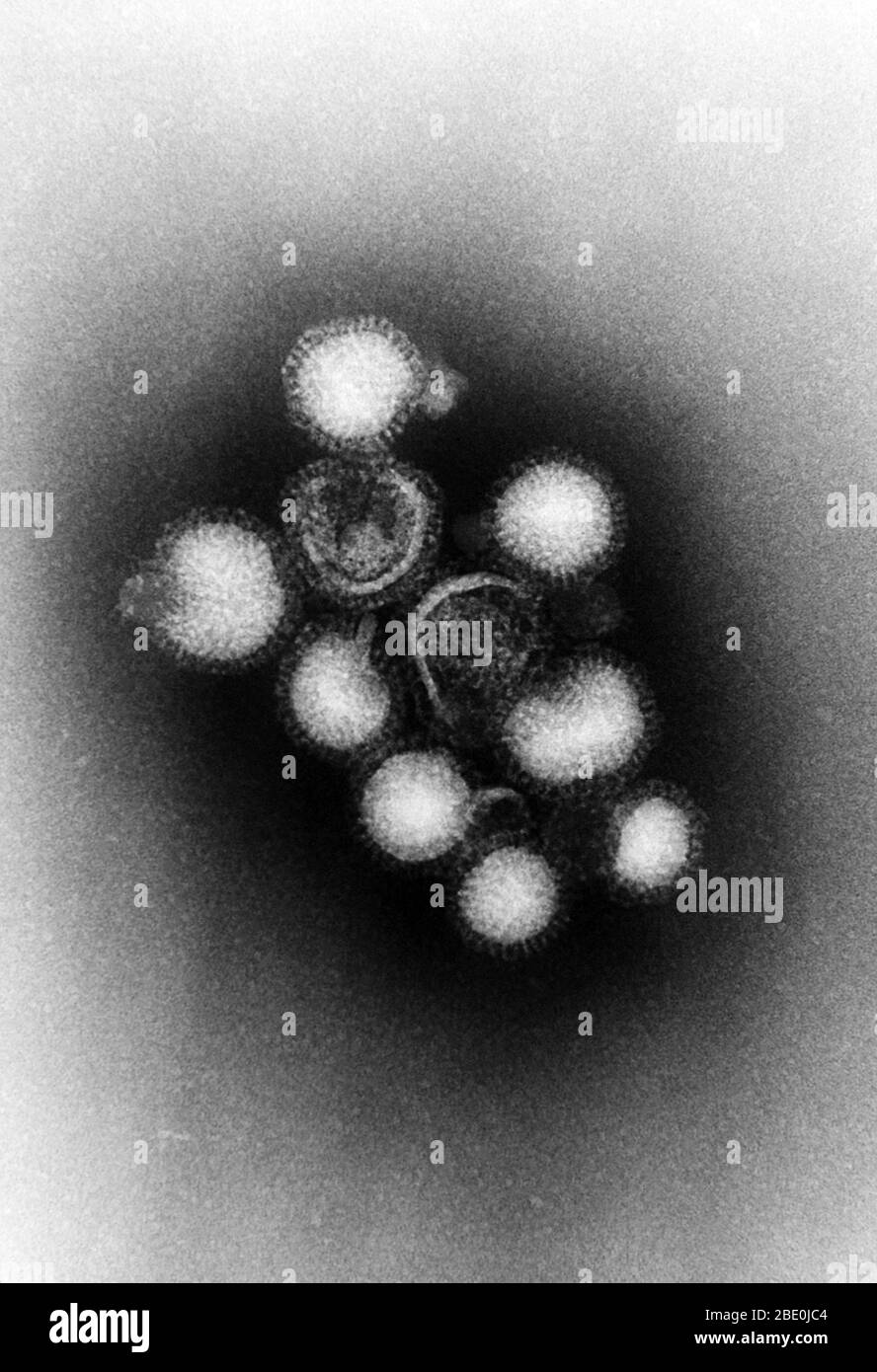
Transmission Electron Microscope (With Diagram) The final image in a TEM is known as transmission electron micrograph. The salts of some heavy metals, e.g., lead; osmium, tungsten and uranium are often used for staining. These heavy metal stains are used to increase the contrast between ultra structures and the background.
Transmission Electron Microscopy - an overview | ScienceDirect Topics Transmission electron microscopy (TEM) TEM was first described in 1932 by Knoll and Ruska, but the resolution of early models was no better than existing light microscopes. TEM resolution exceeded that of light microscopy within three years and, by 1946, resolution to ten Angstroms (1×10 −10 m) had been achieved.

















Post a Comment for "43 transmission electron micrograph labeled"