39 label the glial cells of the cns and pns
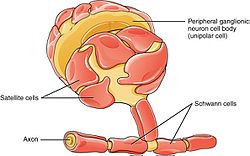
Intact Drosophila central nervous system cellular quantitation reveals ... however, apart from the nervous system of the nematode caenorhabditis elegans (302 neurons, 56 glia) ( white et al., 1986 ), the exact numbers of cells within the central nervous system (cns) of model organisms or that of humans is currently unknown, with estimates, including those based on extrapolation from direct quantification of brain … quizlet.com › 94296903 › glial-cells-of-cns-and-pnsglial cells of CNS and PNS Flashcards | Quizlet oligodendrocytes- glial cells in CNS that have myelin sheath wrap many time around the axon. Schwann cells- glial cells in PNS that have myeline sheath wrap around only one axon. location of satellite cells. suround neuron cell bodies. fuction of satellite of cells.
Cd59 and inflammation regulate Schwann cell development | eLife cd59 is expressed in myelinating glial cells during nervous system development. ( A) Timeline of Schwann cell (SC) (orange) development (top panel). SC developmental stages for zebrafish (hours post fertilization [hpf]) and mice (embryonic day [E]) are indicated in the bottom … Figure 1—source data 1

Label the glial cells of the cns and pns
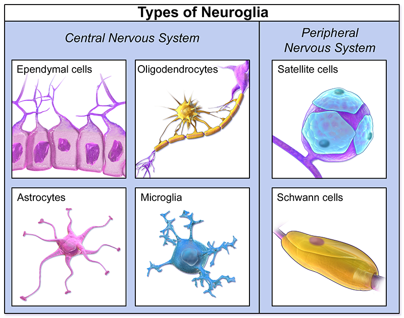
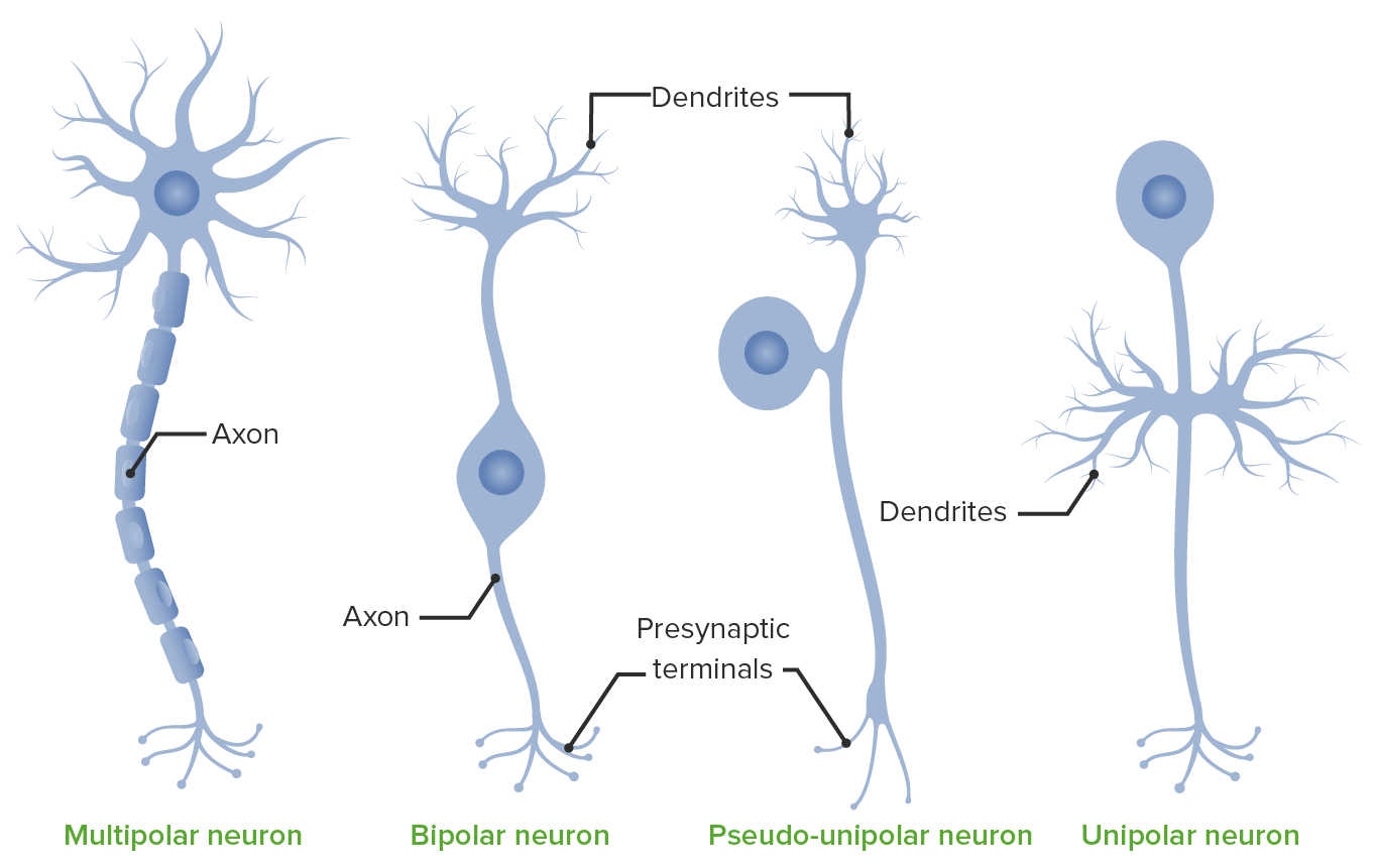
Nervous System: Histology | Concise Medical Knowledge The CNS consists of 4 types of glial cells: oligodendrocytes, astrocytes, microglia, and ependymal cells, each having a different function. In the PNS, the supporting cells are called peripheral neuroglia and include Schwann cells, satellite cells, and various other cells having specific structures and functions. sensory neuron cell body microscope - bobwazneh.com Ganglion = clusters of neuronal cell bodies in the peripheral nervous systems, as well as associated glial cells and axons. There are three classes of neurons: Sensory neurons carry information from the sense organs (such as the eyes and ears) to the brain. B) It is the part of a neuron that carries information to the cell body. › homework-help › questions-andSolved Label the glial cells in the CNS by clicking and ... Expert Answer. ANSWER : Labell …. View the full answer. Transcribed image text: Eved Label the glial cells in the CNS by clicking and dragging the label to the correct location. Myelinated axon Microglial cell Neuron Capillary Oligodendrocyte Cerebrospinal fluid in CNS cavity Astrocyte Ependymal cell Myelin sheath कणा यामम 3 . 7oom.
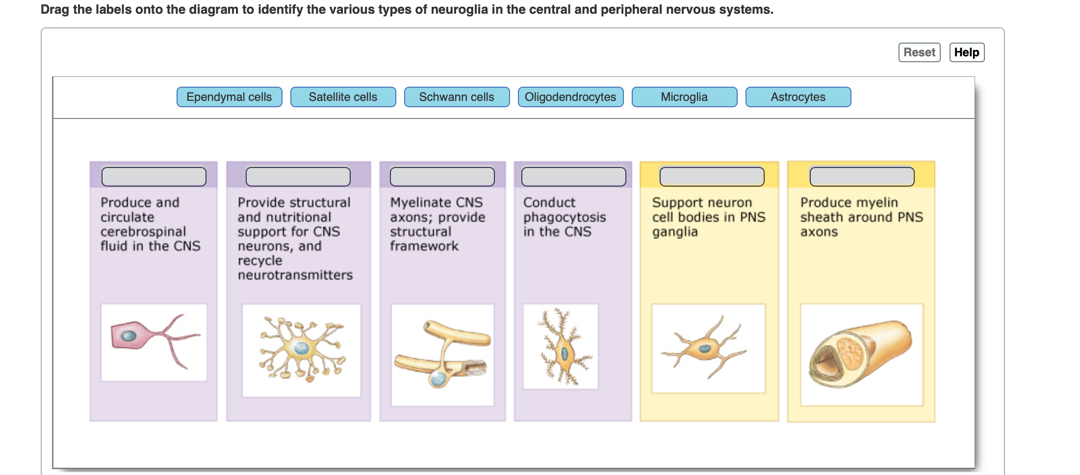
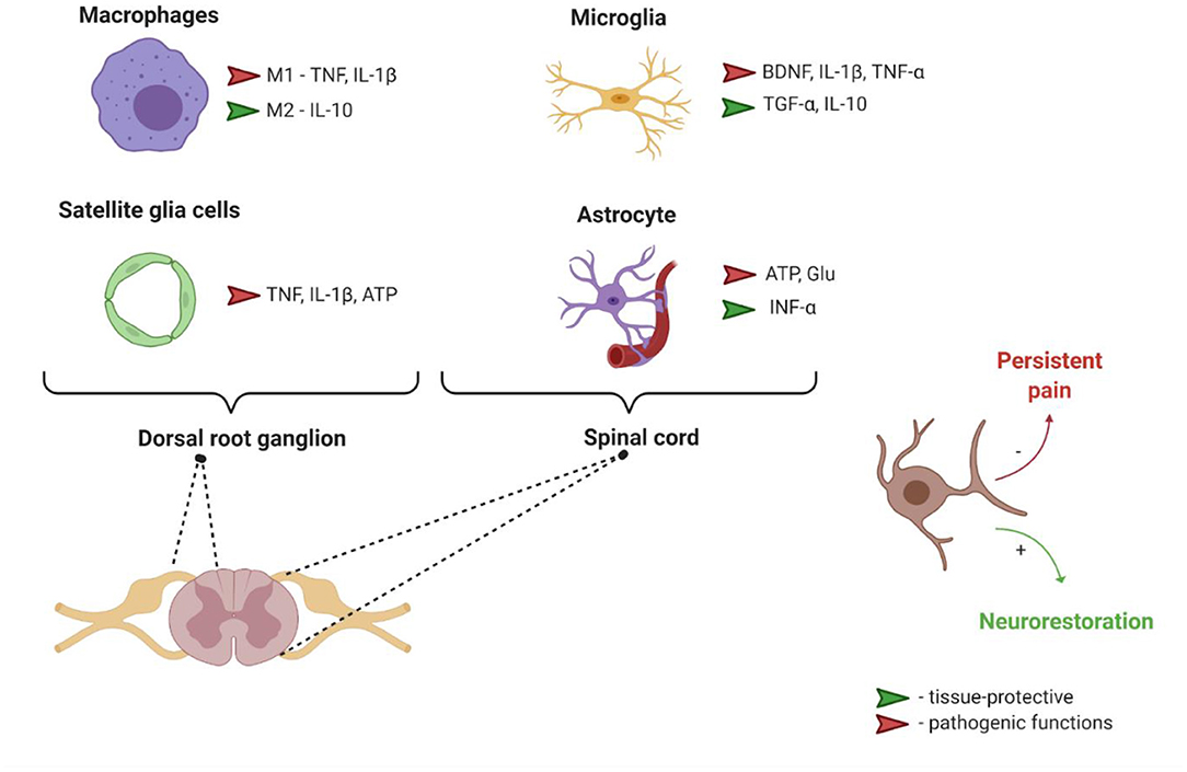
Label the glial cells of the cns and pns. Nervous system: Structure, function and diagram | Kenhub Astrocytes (CNS) and satellite glial cells (PNS) both share the function of supporting and protecting neurons. Other two glial cell types are found in CNS exclusively; microglia are the phagocytes of the CNS and ependymal cells which line the ventricular system of the CNS. Distinct roles of the meningeal layers in CNS autoimmunity Histological analyses of the CNS at the disease peak revealed a strong inflammation of the spinal cord leptomeninges and parenchyma with a massive infiltration of T cells and myeloid cells. In... Autonomic nervous system: Anatomy, divisions, function - Kenhub The autonomic nervous system (ANS) is a functional division of the nervous system, with its structural parts in both the central nervous system (CNS) and the peripheral nervous system (PNS). It controls the glands and smooth muscle of all the internal organs (viscera) unconsciously. This is why it's also called the visceral nervous system. Evaluation of Cytotoxic, Necrotic, Apoptotic, and Autophagic Effects of ... Neuronal cells are typically classified into three types of sensory neurons, motor neurons, and interneurons, while glial cells include oligodendrocytes, astrocytes, ependymal cells, and microglia in the central nervous system and Schwann cells and satellite cells in the peripheral nervous system (Moser et al. 2019).
Extracellular Vesicles From Hyperammonemic Rats Induce ... There are several mechanisms by which peripheral alterations in the immune system may be transmitted to the brain to induce neuroinflammation, including infiltration of peripheral immune cells into the brain (6, 7), activation by peripheral interleukins such as IL-6, IL-1β or IL-17 of their receptors in endothelial cells and transmission of ... Cells of the Blood-Brain Barrier: An Overview of the Neurovascular Unit ... Unlike the endothelial cells lining the peripheral vasculature, brain vascular endothelial cells are completely confluent and interconnected circumferentially by transmembrane tight junction proteins that prevent the passive transfer of cells and molecules between the blood and the brain based on size, surface electrical charge, and lipid ... Nerve cells - ResearchGate The PNS is a different story from the CNS. Rewiring can and does happen in periphery. This type of thing has not been shown convincingly in CNS, except with experimental manipulation of immune... Where labels are aligned in cells? - TimesMojo Right-click and then select "Format Cells" from the popup menu. When the Format Cells window appears, select the Alignment tab. Click on "Center Across Selection" in the drop-down box called Horizontal. Now when you return to your spreadsheet, you should see the text centered across the cells that you selected.
Frontiers | Dysregulation of BMP, Wnt, and Insulin Signaling in Fragile ... Drosophila models of neurological disease contribute tremendously to research progress due to the high conservation of human disease genes, the powerful and sophisticated genetic toolkit, and the rapid generation time. Fragile X syndrome (FXS) is the most prevalent heritable cause of intellectual disability and autism spectrum disorders, and the Drosophila FXS disease model has been critical ... Present and future avenues of cell‐based therapy for brain injury: The ... The latter comprises the main constituents of the central nervous system (CNS), including neurons, interneurons, glia (astrocytes, microglia, and oligodendrocytes), pericytes, endothelial cells, myocytes, and components of the extracellular matrix [ 15 ]. Neurons play a role of "sensors" for variations in the supply of nutrients and oxygen. The Nervous System Anatomy And Physiology Coloring Workbook Answers Oct 14, 2021 · The nervous system subdivides into the central nervous system and the peripheral nervous system. The central nervous system is the brain and spinal cord, while the peripheral nervous system consists of everything else. The central nervous system's responsibilities include receiving, processing, and responding to sensory information. The role of glia in neurological disease - yes, therapy helps! to demonstrate that glia is involved in non-recovery, since it is the only thing that differentiates this neurological alteration when it occurs in snp or in the cns, albert j. aguayo, conducted in the 1980s an experiment in which rats with damaged spinal cord (ie, with paralysis), they received a transplant of the sciatic nerve tissue towards …
Angiotensin type-1 receptor and ACE2 autoantibodies in Parkinson´s ... Introduction. The possible role of autoimmune processes in initiation and/or progression of neurodegeneration, and particularly Parkinson´s disease (PD), has been increasingly suggested 1,2.Different brain disorders, including neurodegenerative diseases, have been associated with autoantibodies that target the extracellular domains of neuronal or glial proteins 3,4.
Tumor-Host Interactions in Malignant Gliomas | SpringerLink The main cell types found in the CNS are neurons, astrocytes, oligodendrocytes, ependymal cells, neural stem cells, progenitor cells, microglia, immune cells, endothelial cells, and pericytes. The extracellular matrix comprises hyaluronic acid, proteoglycans, and glycoproteins and accounts for 10-20% of the volume of the CNS tissue [ 1 ]. Neurons
Imaging of Stem Cell Therapy for Stroke and Beyond Stem cell-based regenerative medicine is an attractive approach for the treatment of neurological diseases such as ischemic stroke. Preclinical studies have shown that transplanted cells can differentiate into neurons and glial cells and improve neurological function. To fully demonstrate and evaluate the potential of stem cell transplantation ...
LDN: A Game Changer for Many Patients - Townsend Letter Complex regional pain syndrome (CRPS) is a neuropathic pain syndrome, which involves glial activation and central sensitization in the central nervous system. The authors describe positive outcomes of two CRPS patients, after they were treated with low-dose naltrexone in combination with other CRPS therapies.
Brain-on-a-Chip | SpringerLink The central nervous system (comprising of the brain and spinal cord) is formed from the inner cells of the neural tube, whereas the peripheral nervous system (comprising nerves outside the brain and spinal cord) is formed by the outer cells. This whole event of the formation of distinct brain structures is called neurulation.
Leukocyte invasion of the brain after peripheral trauma in zebrafish ... Whereas the central nervous system (CNS) is considered immune-privileged from the periphery, the roles of peripheral immune cells remain vital in both homeostasis and disease pathogenesis of the ...
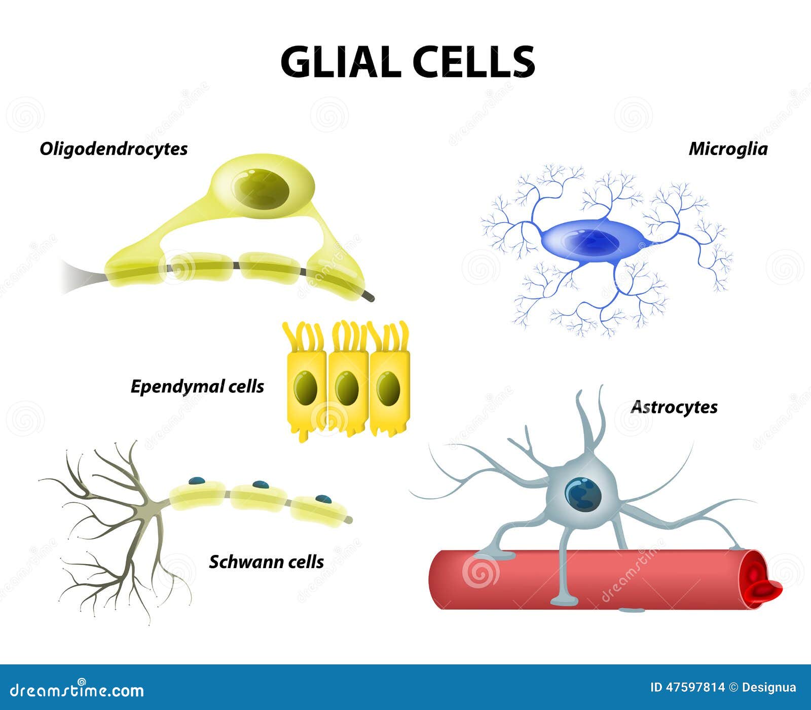
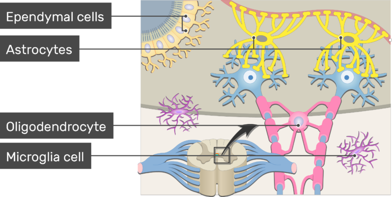
Glial cells: Histology and clinical notes - Kenhub There are four general types of glia in the central nervous system; astrocytes, oligodendrocytes, microglia, and ependymal cells. Some of these cells can be further subdivided based on their embryology. Oligodendrocytes Oligodendrocytes have long cytoplasmic projections extending from their soma.
Schwann cells: Anatomy, function and histology | Kenhub The Schwann cells, also known as neurolemmocytes, are a type of glial cells present exclusively in the peripheral nervous system. They develop from precursors in the neural crest and can be differentiated into two types of cells: Myelinating Schwann cells Nonmyelinating Schwann cells
Glial Cells - Neuroanatomy Basics - BYU-Idaho In this anatomy tutorial we look at the different types of glial cells in the central and peripheral nervous system. The following structures are discussed: - astrocytes, vascular foot process. - oligodendrocytes, myelin/myelination, - microglial cell. - ependymal cells, cilia, brain ventricles, choroid plexus, cerebrospinal fluid.
Frontiers | Uncovering the spectrum of adult zebrafish neural stem cell ... Adult neural stem and progenitor cells (aNSPCs) persist lifelong in teleost models in diverse stem cell niches of the brain and spinal cord. Fish maintain developmental stem cell populations throughout life, including both neuro-epithelial cells (NECs) and radial-glial cells (RGCs). Within stem cell domains of the brain, RGCs persist in a cycling or quiescent state, whereas NECs continuously ...
› glial-cellsGlial Cells Types and Functions - Simply Psychology Jul 08, 2021 · Glial cells, also called glial cells or neuroglia, are cell which are non-neuronal and are located within the central nervous system and the peripheral nervous system that provides physical and metabolic support to neurons, including neuronal insulation and communication, and nutrient and waste transport. Glial cells are a general term for many types of glial cell, for example microglial, astrocytes, and Schwann cells, each having their own functions within the body.
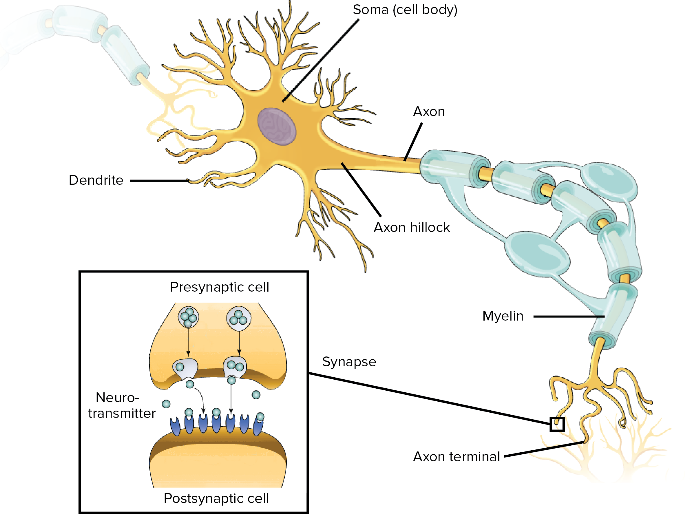
Brain, spinal cord and peripheral nervous system anatomy | Kenhub Myelin insulates axons and allows quicker transmission of electrical impulses. A bundle of axons (nerve fibers) in the CNS is called a tract. In the PNS this bundle is called a nerve. Besides from neurons, there are other nervous system cells, for example glial cells, which play supporting roles. Learn about the cells of the nervous system here.
Nervous Tissue Structure to its location: 1. CNS: located and protected by the cranial cavity (brain), the vertebral canal (spinal cord). 2. PNS: any nervous tissue outside the brain or the spinal cord. Anatomy and Physiology of - Jones & Bartlett Learning A solid, wall-like structure that separates the left atria . and ventricle from the right atria and ventricle.
› homework-help › questions-andSolved Label the glial cells of the CNS and PNS. Astrocyte ... We review their content and use your feedback to keep the quality high. Answer ; Glial cells, also known as neuroglia, are non-neuronal cells that offer physical …. View the full answer. Transcribed image text: Label the glial cells of the CNS and PNS. Astrocyte Satellite cell Microglial cell Eperdymal cell Oligodendrocyte Schwann cell.
quizlet.com › 134481702 › glial-cells-of-the-cns-andGlial Cells of the CNS and PNS Flashcards | Quizlet Start studying Glial Cells of the CNS and PNS. Learn vocabulary, terms, and more with flashcards, games, and other study tools.
› homework-help › questions-andSolved Label the glial cells in the CNS by clicking and ... Expert Answer. ANSWER : Labell …. View the full answer. Transcribed image text: Eved Label the glial cells in the CNS by clicking and dragging the label to the correct location. Myelinated axon Microglial cell Neuron Capillary Oligodendrocyte Cerebrospinal fluid in CNS cavity Astrocyte Ependymal cell Myelin sheath कणा यामम 3 . 7oom.
sensory neuron cell body microscope - bobwazneh.com Ganglion = clusters of neuronal cell bodies in the peripheral nervous systems, as well as associated glial cells and axons. There are three classes of neurons: Sensory neurons carry information from the sense organs (such as the eyes and ears) to the brain. B) It is the part of a neuron that carries information to the cell body.
Nervous System: Histology | Concise Medical Knowledge The CNS consists of 4 types of glial cells: oligodendrocytes, astrocytes, microglia, and ependymal cells, each having a different function. In the PNS, the supporting cells are called peripheral neuroglia and include Schwann cells, satellite cells, and various other cells having specific structures and functions.







:max_bytes(150000):strip_icc()/glialcellsillustration-5a94d585642dca00362568c4.jpg)





:background_color(FFFFFF):format(jpeg)/images/library/2394/a6Zzn18njB3aIQgWosmC3Q_Myelin.png)
Post a Comment for "39 label the glial cells of the cns and pns"